
Histomorphological patterns and Helicobacter pylori status in gastric tissue biopsies assessed by Giemsa histochemical staining: a hospital-based cross-sectional study at Bugando Medical Centre, Mwanza, Tanzania
Mary Felician Shayo, Oscar Ottoman Muhini, Edrick Elias Mutalemwa, Johnson Paul Nyansuku, Ester Reuben Bugumba, Hyasinta Jaka Jaka
Corresponding author: Oscar Ottoman Muhini, Pathology Department, Catholic University of Health and Allied Sciences, Weill Bugando School of Medicine, Mwanza, Tanzania 
Received: 18 Feb 2023 - Accepted: 18 Jun 2026 - Published: 18 Aug 2026
Domain: Anatomic pathology, Gastroenterology
Keywords: Histomorphological patterns, Helicobacter pylori, Giemsa stain
Funding: This work received no specific grant from any funding agency in the public, commercial, or non-profit sectors.
©Mary Felician Shayo et al. Pan African Medical Journal (ISSN: 1937-8688). This is an Open Access article distributed under the terms of the Creative Commons Attribution International 4.0 License (https://creativecommons.org/licenses/by/4.0/), which permits unrestricted use, distribution, and reproduction in any medium, provided the original work is properly cited.
Cite this article: Mary Felician Shayo et al. Histomorphological patterns and Helicobacter pylori status in gastric tissue biopsies assessed by Giemsa histochemical staining: a hospital-based cross-sectional study at Bugando Medical Centre, Mwanza, Tanzania. Pan African Medical Journal. 2026;54:126. [doi: 10.11604/pamj.2026.54.126.39339]
Available online at: https://www.panafrican-med-journal.com//content/article/54/126/full
 PDF in process
 Research 

Histomorphological patterns and Helicobacter pylori status in gastric tissue biopsies assessed by Giemsa histochemical staining: a hospital-based cross-sectional study at Bugando Medical Centre, Mwanza, Tanzania
Histomorphological patterns and Helicobacter pylori status in gastric tissue biopsies assessed by Giemsa histochemical staining: a hospital-based cross-sectional study at Bugando Medical Centre, Mwanza, Tanzania
Mary Felician Shayo1,  Oscar Ottoman Muhini1,2,&, Edrick Elias Mutalemwa1,2, Johnson Paul Nyansuku2,3, Ester Reuben Bugumba2, Hyasinta Jaka Jaka4
&Corresponding author
Introduction: Helicobacter pylori (H. pylori) infection is a leading cause of chronic gastric disease and malignancy worldwide. In resource-limited settings, accurate detection is essential for clinical management. This study evaluated histomorphological patterns of gastric lesions and H. pylori status among patients at Bugando Medical Centre (BMC) in Mwanza, Tanzania.
Methods: a hospital-based cross-sectional study was conducted using 300 archival gastric tissue biopsy specimens. Histomorphological assessment was performed using haematoxylin and eosin staining. H. pylori status was determined using a Giemsa histochemical stain. Data were analysed using SPSS version 20, with statistical significance set at p <0.05.
Results: males constituted 161/300 (53.7%) of the cohort, and 202/300 (67.3%) of participants were aged 40 years. The most frequent histomorphological diagnoses were chronic gastritis (131/300, 43.7%) and gastric adenocarcinoma (115/300, 38.3%). The overall prevalence of H. pylori was 35.3%. Positivity was significantly higher in males (52.8%) than in females (15.1%, p=0.001) and in patients aged <20 years (100.0%). H. pylori was detected in 90.0% of gastric ulcers and 100.0% of gastric perforations. Multivariate analysis identified male sex (aOR=5.64; 95% CI: 2.41-13.22) and younger age (aOR=0.37 for age <40 years) as independent predictors of infection.
Conclusion: chronic gastritis and adenocarcinoma are the predominant gastric pathologies at BMC. The high H. pylori burden, particularly among younger males and patients with ulcerative disease, underscores the need for routine Giemsa staining to improve diagnostic accuracy and guide the selection of effective eradication therapy in northwestern Tanzania.

Helicobacter pylori is a small, curved, highly motile Gram-negative bacillus that colonises the gastric mucosa [1]. The organism is primarily transmitted via the faecal-oral route through contaminated food and water, and it infects approximately 50% of the global population [1,2]. The burden is particularly high in developing countries, where poor sanitation and limited access to clean water facilitate transmission[2]. In Tanzania, recent meta-analyses estimate a pooled prevalence of 54.3%, making it a major public health concern [3].
Helicobacter pylori infection is associated with a wide range of gastric diseases, including acute and chronic gastritis, peptic ulcer disease, and gastric malignancies [3]. Persistent infection causes chronic gastric inflammation that may progress through atrophic gastritis, intestinal metaplasia, dysplasia, and ultimately to gastric adenocarcinoma [1]. A multinational study of populations from 13 countries found that infected individuals had approximately six times the risk of gastric cancer compared with uninfected individuals [4]. Studies across Africa have also documented high rates of H. pylori infection among patients with gastric cancer and other gastric lesions, underscoring its important role in gastric carcinogenesis [5].
The prevalence of H. pylori varies geographically, with a global prevalence of 44.3% [5]. Africa bears the highest burden worldwide, with prevalence approaching 80% in some populations [6]. In sub-Saharan Africa, up to 50% of children acquire the infection by age 5 [7]. In Tanzania, prevalence ranges from 50% in the general population to over 65% among patients with dyspeptic symptoms in some regions [3,8]. Despite this high burden, information on the histomorphological spectrum of gastric lesions and the distribution of H. pylori infection in gastric biopsy specimens from Mwanza, Northwestern Tanzania, remains limited [3]. Several methods are available for detecting H. pylori in gastric biopsies, including haematoxylin and eosin (H&E), modified Giemsa staining, and immunohistochemistry [9]. Although immunohistochemistry is regarded as the reference standard for its high sensitivity (98%), its routine use is limited in resource-constrained settings due to cost and technical requirements [10]. Modified Giemsa staining is a simple, affordable, and reliable alternative with superior diagnostic performance compared with routine H&E staining [9,10].
At Bugando Medical Centre, gastric biopsy diagnosis relies mainly on H&E staining, which may lead to underdiagnosis of infection [11]. Therefore, this study aimed to determine the histomorphological patterns of gastric lesions and to assess H. pylori status using Giemsa staining in gastric biopsy specimens received at Bugando Medical Centre.

Study design and study setting: this was a retrospective cross-sectional laboratory-based study conducted between April and July 2022 at the Histopathology Unit of the Central Pathology Laboratory, Bugando Medical Centre (BMC), Mwanza, Tanzania. The study involved reviewing archived gastric tissue biopsy paraffin blocks collected between January 2020 and January 2022. BMC serves as a tertiary zonal referral and teaching hospital for the Catholic University of Health and Allied Sciences (CUHAS). BMC serves the northwestern region of Tanzania with a catchment area of approximately 20 million people across eight administrative regions. BMC receives approximately 7000 tissue biopsies and 2500 cytopathological samples for pathological diagnosis annually.
Study population and sampling strategy: the study included archived gastric tissue biopsy paraffin blocks retrieved from the Histopathology Unit of the Central Pathology Laboratory at Bugando Medical Centre. Eligible specimens were all gastric tissue biopsy blocks archived between January 2020 and January 2022. Tissue blocks that were lost, contained no tissue sections, or were inadequate for histopathological diagnosis were excluded from the study.
The minimum sample size was calculated using the Leslie Kish formula, based on a previously reported prevalence of Helicobacter pylori of 37.1%, detected by H. pylori antibody immunohistochemical staining in a study conducted at Muhimbili National Hospital [12], and a standard error of 0.05. The sample size (N) is calculated using the formula below:

Where Z = standard normal deviate corresponding to a 95% confidence level (1.96); P = estimated prevalence of Helicobacter pylori (0.371); and d = desired precision (0.05). Substituting the values into the formula:

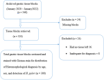
Thus, the minimum calculated sample size was approximately 359 gastric tissue biopsy specimens. However, a total of 348 eligible gastric tissue biopsy blocks archived between January 2020 and January 2022 were available and included in the study. All eligible specimens were included in the study using a total sampling approach (Figure 1).
Data collection procedure and gastric tissue block recruitment: of the 348 documented gastric tissue blocks, only 300 met the inclusion criteria and were recruited. Twenty-four were excluded due to missing blocks, and another 16 cases had no tissue remaining. 8 were inadequate for diagnosis due to poor morphology in tissue sections (Figure 1). Demographics and clinical factors were retrieved from the medical records and the laboratory DISA system and documented in a structured checklist. In this study, the independent variable assessed was H. pylori status, while the dependent variables were age groups, sex, and histomorphological diagnosis.
Hematoxylin and eosin laboratory procedure: the retrieved tissue blocks were cut into thin sections (2-3 μm) using a Rotary Microtome. The tissues were then stained with routine hematoxylin and eosin (H&E). Deparaffinization was performed in a hot-air oven at 60°C, and the slides were then placed in two xylene containers for 10 minutes each. This was followed by rehydration, in which the tissue slides were dipped into a series of absolute ethanol (99.8%) and 95% ethanol for 10 minutes, then into clean tap water for 5 minutes. The tissue slides were then immersed in hematoxylin for 10 minutes and then blued in clean running tap water for 5 minutes. The section was immersed in eosin stain solution for 5 minutes, then dehydrated in 95% and 99.8% ethanol for 10 minutes each. After this, the tissue slides were immersed 10 times in two containers of xylene to clear the ethanol [13,14]. Lastly, the slides were mounted using dibutyl phthalate polystyrene xylene (DPX) for histological diagnosis [14].
Giemsa histochemical staining laboratory procedure: formalin-fixed, paraffin-embedded gastric tissue blocks were sectioned at 2-3 μm thickness and mounted on glass slides. Sections were deparaffinised in xylene, rehydrated through graded alcohols to distilled water, and stained using the Giemsa histochemical method. After staining, slides were rinsed, dehydrated through ascending alcohol concentrations, cleared in xylene, and mounted with coverslips for microscopic examination [15,16].
The stained sections were examined under light microscopy for the presence of Helicobacter pylori. The organisms were identified as dark blue, curved or spiral-shaped bacilli on the gastric surface epithelium and within the mucus layer, against a lightly stained background [15].
The researcher and two registered pathologists independently examined the stained slides for H&E and Giemsa under an Olympus CX21 light microscope for pathological tissue diagnosis and evaluation of Helicobacter pylori staining status [14,16]. Histomorphological diagnoses and agreement on Helicobacter pylori status were recorded, and all Giemsa-stained slides were assessed using known positive control slides. All findings were recorded using a standardized data checklist.
Statistical analysis: data were analyzed using Stata version 17. Categorical variables were summarized using frequencies and percentages. Associations between Helicobacter pylori status and independent variables were assessed using Pearson's chi-square test or Fisher's exact test, as appropriate. Clinically relevant variables were included in binary logistic regression to estimate crude and adjusted odds ratios with 95% confidence intervals. A p-value of <0.05 was considered statistically significant.
Ethical approval and ethical considerations: ethical clearance was granted by the joint CUHAS/BMC Research and Ethics Review Committee, with undergraduate ethical clearance certificate No. 2324/2022. Administrative permission to conduct the study was obtained from Bugando Medical Centre through the head of the Histopathology Unit.
As this was a retrospective study involving archived tissue specimens, patient confidentiality was strictly maintained. No personal identifiers were collected. Data were entered into a password-protected database accessible only to the research team.
Study population characteristics: in this study, 300 gastric tissue biopsy specimens were included in the analysis. Males accounted for 53.7% (n = 161) of cases, while females constituted 46.3% (n = 139). The majority of patients were aged ≥40 years (67.3%, n = 202), followed by those aged 20-39 years (24.7%, n = 74) (Table 1). Histomorphological assessment showed that chronic gastritis was the most frequent diagnosis, representing 43.7% (n = 131) of cases, followed by gastric adenocarcinoma (38.3%, n = 115) (Table 1, Figure 2).
Among 300 gastric tissue biopsy specimens included in this study, Helicobacter pylori was detected in 35.3% (n = 106), while 64.7% (n = 194) were negative.
Association between Helicobacter pylori infection and clinicopathological variables: significant associations were observed between H. pylori status and sex, age group, and histomorphological diagnosis (p <0.05 for all comparisons). Males had a higher proportion of H. pylori-positive cases (52.8%) than females (15.1%). Age stratification showed a decreasing trend in infection prevalence with increasing age. All participants aged <20 years were H. pylori-positive (100.0%), whereas those aged ≥40 years had the highest proportion of negative cases (76.7%). Histomorphological findings revealed variation in the distribution of infection across lesion types. Higher proportions of H. pylori positivity were observed in gastric ulcer (90.0%) and gastric perforation (100.0%), whereas chronic gastritis showed 45.0% positivity. Lower proportions were observed in gastric adenocarcinoma (22.6%), gastric polyps (11.1%), and gastrointestinal stromal tumor (GIST) (16.7%). Normal gastric mucosa showed 57.1% positivity (Table 2, Figure 3).
Univariate analysis of factors associated with H. pylori infection: in univariate logistic regression, male sex was strongly associated with increased odds of H. pylori infection (cOR = 6.28, 95% CI: 3.60-10.98). Participants aged ≥40 years had significantly lower odds of infection than those aged <40 years (cOR = 0.20, 95% CI: 0.12-0.34) (Table 3). Histomorphological diagnosis also showed significant variation. Compared with normal gastric mucosa, gastric adenocarcinoma and benign lesions were associated with reduced odds of infection, whereas gastritis and ulcerative lesions showed variable but generally lower crude odds of infection. Overall, lesion type showed significant heterogeneity in the distribution of infection.
Multivariate analysis of independent predictors of H. pylori infection: after adjustment for potential confounders, sex, age, and histomorphological diagnosis remained independently associated with H. pylori infection. Male sex was associated with more than fivefold higher odds of infection than female sex (aOR = 5.64, 95% CI: 2.41-13.22). Participants aged ≥40 years had significantly lower odds of infection than younger individuals (aOR = 0.37, 95% CI: 0.17-0.80). Compared with normal gastric mucosa, gastric adenocarcinoma, benign lesions, and gastritis/ulcerative conditions were all associated with lower odds of H. pylori detection, with the strongest inverse association observed in the gastritis/ulcer group (aOR = 0.07, 95% CI: 0.01-0.44) (Table 3).
The findings of this study offer critical insights into the histomorphological spectrum of gastric lesions and the prevalence of Helicobacter pylori at Bugando Medical Centre, a tertiary referral hospital in Mwanza, Tanzania. Analysing 300 gastric biopsies, the study identifies key demographic and pathological associations that are essential for improving local diagnostic and therapeutic strategies.
In this study, the prevalence of Helicobacter pylori detected by Giemsa staining was 35.3%, which is lower than the national pooled prevalence but consistent with the declining trend reported in Tanzania [3,8]. Chronic gastritis was the most common histomorphological diagnosis, followed by gastric adenocarcinoma, reflecting the substantial burden of gastric inflammatory and malignant diseases at this tertiary referral hospital. Male sex and younger age were significantly associated with H. pylori infection, suggesting early acquisition of the organism and higher susceptibility among males. H. pylori positivity was particularly high among patients with gastric ulcers and perforations, supporting its important role in peptic ulcer disease. Giemsa staining improves the detection of H. pylori compared with routine H&E staining and provides a practical, cost-effective diagnostic alternative in resource-limited settings [9,10].
Prevalence of H. pylori infection: the overall detection rate of H. pylori using Giemsa staining was 35.3%. This finding is notably lower than the pooled national prevalence of 54.3% reported in a recent systematic review and meta-analysis of Tanzanian studies [3]. However, our results align with a documented decline in H. pylori prevalence in Tanzania, from approximately 79% in earlier studies to around 36% in more recent years [3]. This variation may also reflect regional differences; prior seroprevalence data from the Mwanza region reported a rate of 39.1%, suggesting that the Lake Zone may have lower infection densities than other parts of the country [3].
Histomorphological spectrum of gastric lesions: the most frequent histomorphological finding was chronic gastritis, followed closely by gastric adenocarcinoma. This high proportion of gastritis aligns with other reports from a study conducted in the Lake Zone area of Western Tanzania, where chronic inflammation remains the leading indication for gastric biopsy [11].
The high frequency of gastric adenocarcinoma (38.3%) observed in this study is particularly notable compared with other African settings, such as Cameroon, where cancer rates in biopsy cohorts were lower [17]. This high burden of malignancy likely reflects the selection bias inherent in a tertiary centre like BMC; patients often present with advanced symptoms such as significant weight loss or haematemesis, which are late manifestations of gastric cancer [11].
Factors associated with H. pylori infection: our study identified male sex as a strong independent predictor of H. pylori infection. Males had a significantly higher positivity rate than females, and multivariate analysis showed that they had more than fivefold higher odds of infection. This disparity may be linked to differences in health-seeking behaviours or environmental exposures in the Lake Zone, where males are often diagnosed at more advanced stages of gastric disease [3,11]. Regarding age, we observed a 100% positivity rate among participants under 20 years of age, with infection rates decreasing with increasing age. This pattern supports the early acquisition hypothesis, in which H. pylori is acquired in early childhood in developing countries due to factors such as overcrowding and poor water quality [7,18]. The lower detection rates in older patients may reflect progression of gastritis to atrophic stages, creating a hostile, hypoacidic environment that promotes the natural clearance of the bacteria [3].
Association with ulcerative and malignant lesions: the distribution of infection varied markedly across lesion types. We found a very strong association between gastric ulcers and gastric perforations, confirming H. pylori as a major driver of peptic ulcer disease in this population [3]. By contrast, only 22.6% of patients with gastric adenocarcinoma were H. pylori-positive. Although the infection is a known risk factor for cancer development through the Correa cascade (gastritis to metaplasia to cancer), the bacteria often disappear once malignancy is established due to the altered gastric microenvironment [2].
Diagnostic utility of Giemsa staining: the results underscore the value of Giemsa staining in routine pathology practice. A study conducted in Tanzania reported that routine hematoxylin and eosin (H&E) staining has historically yielded a low detection rate of approximately 11.2% [9,12]. In our study, Giemsa staining increased the detection rate to 35.3%, demonstrating its superior sensitivity for detecting the organism [9]. In resource-limited settings such as Tanzania, where immunohistochemistry (the gold standard) is often unavailable due to cost, Giemsa provides a cost-effective and reliable alternative that supports more accurate clinical management [9,10].
This study analyzed a relatively large sample of 300 gastric tissue biopsies from a tertiary referral hospital, providing valuable data on the histomorphological spectrum of gastric lesions and Helicobacter pylori infection in northwestern Tanzania. Giemsa histochemical staining improved H. pylori detection compared with routine haematoxylin and eosin staining, demonstrating its utility as a cost-effective diagnostic method in resource-limited settings. In addition, independent slide review by two pathologists and multivariable logistic regression strengthened the reliability of the findings. However, the retrospective cross-sectional design limits causal inference and may be subject to selection bias, as only archived tissue blocks with adequate tissue and complete records were included. The absence of immunohistochemical confirmation may have led to an underestimation of H. pylori prevalence. At the same time, the use of archival biopsy specimens with limited tissue sampling may have reduced detection sensitivity. Furthermore, important clinical and behavioural variables, including endoscopic findings, symptom severity, prior antibiotic or proton-pump inhibitor use, smoking, alcohol consumption, and socioeconomic factors, were unavailable due to the retrospective study and the lack of that information in the Electronic Data Hospital Management System (EHMS). Interobserver agreement between two pathologists was not calculated statistically during the study and is acknowledged as a limitation of this study.
Helicobacter pylori infection remains a significant health burden at Bugando Medical Centre, particularly among younger males and patients with ulcerative conditions. The high prevalence of gastric adenocarcinoma in this region warrants early screening and routine use of Giemsa staining as a cost-effective diagnostic tool to improve H. pylori detection and enable timely eradication therapy.
What is known about this topic
- Helicobacter pylori causes chronic gastritis, peptic ulcer disease, and gastric cancer, necessitating accurate diagnosis for effective management;
- Histopathological examination of gastric biopsies remains important for diagnosing H. pylori infection and for evaluating gastric lesions;
- Giemsa staining improves H. pylori detection, but epidemiological and histomorphological patterns remain poorly characterised in sub-Saharan Africa.
What this study adds
- Chronic gastritis and gastric adenocarcinoma were the most common histomorphological diagnoses in gastric biopsies at Bugando Medical Centre;
- Over one-third of patients had H. pylori infection, strongly associated with gastric ulceration and perforation;
- Male sex and younger age predicted H. pylori infection; routine Giemsa staining improved detection accuracy.
The authors declare no competing interests.
Oscar Ottoman Muhini, Mary Felician Shayo, Edrick Elias Mutalemwa, and Hyasinta Jaka Jaka conceived, designed, and coordinated the study; Mary Felician Shayo and Ester Reuben Bugumba performed the histopathological laboratory bench work; Oscar Ottoman Muhini and Edrick Elias Mutalemwa reviewed slides for pathological diagnosis and H. pylori status; Johnson Paul Nyansuku conducted the statistical analysis; Oscar Ottoman Muhini, Mary Felician Shayo, and Hyasinta Jaka Jaka wrote the initial draft of the manuscript, which all authors critically revised; Oscar Ottoman Muhini is the guarantor of the study. All the authors read and approved the final version of this manuscript.
The authors wish to acknowledge the support of the members of the Histopathology and Morbid Anatomy and Internal Medicine-Gastroenterology Units at BMC, the Department of Pathology at CUHAS, and all study participants. We also sincerely thank the Catholic University of Health and Allied Science for assisting with the completion of Mary Felician Shayo's Bachelor of Medical Laboratory Science through this dissertation study.
Table 1: demographic, histomorphological, and Helicobacter pylori characteristics of patients with gastric tissue biopsies assessed at Bugando Medical Centre, Mwanza, Tanzania, from January 2020 to January 2022 (N=300)
Table 2: association between Helicobacter pylori status and demographic and histomorphological characteristics among patients with gastric tissue biopsies assessed at Bugando Medical Centre, Mwanza, Tanzania, from January 2020 to January 2022 (N=300)
Table 3: univariate and multivariate logistic regression analysis of demographic and histomorphological factors associated with Helicobacter pylori positivity among patients with gastric tissue biopsies assessed at Bugando Medical Centre, Mwanza, Tanzania, from January 2020 to January 2022 (N=300)
Figure 1: flow diagram showing the selection and processing of archived gastric tissue blocks for histomorphological assessment and detection of Helicobacter pylori using Giemsa histochemical staining at Bugando Medical Centre, Mwanza, Tanzania, from January 2020 to January 2022 (N=348)
Figure 2: hematoxylin and eosin (H&E) gastric tissue section with various histomorphological diagnoses: (A) gastric ulcers pointed by a black arrow (x10 hpf); (B) chronic gastritis with numerous lymphocytes and plasma cells accumulation pointed by the black arrow; (C) infiltrating gastric adenocarcinoma of intestinal type pointed by the black arrow (x10 hpf)
Figure 3: (A,B) Giemsa-stained gastric mucosa showing Helicobacter pylori organisms within the gastric pits (black arrow), identified as darkly staining curved or bacillary organisms (x40 hpf)
- Ford AC, Yuan Y, Moayyedi P. Helicobacter pylori eradication therapy to prevent gastric cancer: systematic review and meta-analysis. Gut. 2020 Dec;69(12):2113-2121. PubMed | Google Scholar
- Wroblewski LE, Peek RM Jr, Wilson KT. Helicobacter pylori and gastric cancer: factors that modulate disease risk. Clin Microbiol Rev. 2010 Oct;23(4):713-39. PubMed | Google Scholar
- Jaka H, Kilonzo SB, Nkandala I, Rudovick L, Luchius I, Kajogoo VD et al. Prevalence, risk factors, and endoscopic findings among individuals infected with Helicobacter pylori in Tanzania: A systematic review and meta-analysis. BMC Infect Dis. 2026 Feb 14;26(1):598. PubMed | Google Scholar
- THE EUROGAST study group. An international association between Helicobacter pylori infection and gastric cancer. The EUROGAST Study Group. Lancet. 1993 May 29;341(8857):1359-62. PubMed | Google Scholar
- Collatuzzo G, Dajti E, Secco M, Bazzoli F, Boffetta P, Zagari RM. Burden of gastric cancer attributable to Helicobacter pylori in 27 countries from seven geographic regions in 2022. Gastric Cancer. 2026 Jan;29(1):16-26. PubMed | Google Scholar
- Goh KL, Chan WK, Shiota S, Yamaoka Y. Epidemiology of Helicobacter pylori infection and public health implications. Helicobacter. 2011 Sep;16 Suppl 1(0 1):1-9. PubMed | Google Scholar
- Bardhan PK. Epidemiological features of Helicobacter pylori infection in developing countries. Clin Infect Dis. 1997 Nov;25(5):973-8. PubMed | Google Scholar
- Jaka H, Mueller A, E Mshana S, Kasang C. Prevalence and factors associated with active H. pylori infection among the asymptomatic population and dyspeptic patients in Mwanza North-western Tanzania. Acta Scientific Gastrointestinal Disorders. 2022 Jun 1;25-33.
- Alwahaibi N, Al Mamari AM, Al Aamri A, Al Sulimani Y, Al Balushi A. Assessing the diagnostic accuracy of routine hematoxylin and eosin, Alcian Blue/Periodic Acid-Schiff, and Giemsa stains in the detection of Helicobacter pylori in gastric biopsies. F1000Res. 2026 Jan 10;14:1148. PubMed | Google Scholar
- Alturkistani HA, Tashkandi FM, Mohammedsaleh ZM. Histological Stains: A Literature Review and Case Study. Glob J Health Sci. 2015 Jun 25;8(3):72-9. PubMed | Google Scholar
- Munema F, Rambau P, Byabato S, Binde A, Mabega N. Endoscopic and Clinicopathological Patterns of Upper Gastrointestinal Conditions Among Patients Receiving Endoscopic Examinations at Bugando Medical Centre, Mwanza, Tanzania. European Journal of Preventive Medicine. 2025 Mar 18;13(2):34-42.
- Ngaiza AI, Yahaya JJ, Mwakimonga AK, Vuhahula E, Mnango L, Mwakigonja AR et al. Histologic detection of Helicobacter pylori by the immunohistochemical method using anti-Helicobacter pylori polyclonal antibody: A cross-sectional study of patients with gastric pathologies at the Muhimbili National Hospital in Dar-es-salaam, Tanzania. Arab J Gastroenterol. 2022 Feb;23(1):7-14. PubMed | Google Scholar
- Alemany L, Saunier M, Alvarado-Cabrero I, Quirós B, Salmeron J, Shin HR et al. Human papillomavirus DNA prevalence and type distribution in anal carcinomas worldwide. Int J Cancer. 2015 Jan 1;136(1):98-107. PubMed | Google Scholar
- Ottoman O, David F, Nyansuku J, James M, Minja CA, Mukerebe C et al. Genotypes of human papillomavirus and clinical pathological features among patients with anal carcinoma in North-western Tanzania. Infect Agent Cancer. 2026 Apr 1;21(1):37. PubMed | Google Scholar
- Grey SF, Wyatt JI, Rathbone BJ. Simplified techniques for identifying Campylobacter pylori. J Clin Pathol. 1986 Nov;39(11):1279.1. Google Scholar
- Kocsmár É, Szirtes I, Kramer Z, Szijártó A, Bene L, Buzás GM et al. Sensitivity of Helicobacter pylori detection by Giemsa staining is poor in comparison with immunohistochemistry and fluorescent in situ hybridization and strongly depends on inflammatory activity. Helicobacter. 2017 Aug;22(4). PubMed | Google Scholar
- Florice F, Danny T, Ngemeshe S, Paul T, Moungo-Ndjole C, Michele T. Prevalence of Helicobacter pylori and histopathological features of gastric biopsies from patients with dyspeptic symptoms in cameroon: A contraindication of african enigma. Int J Biol. 2025;17(1):1. Google Scholar
- Khalifa MM, Sharaf RR, Aziz RK. Helicobacter pylori: a poor man's gut pathogen? Gut Pathog. 2010 Dec 31;2(1):2. PubMed | Google Scholar
Search
This article authors
On Pubmed
On Google Scholar
Citation [Download]
Navigate this article
Similar articles in
Key words
Tables and figures



Article metrics
Recently from the PAMJ






Authors´ services
