
Risk factors for postoperative palatal fistula following repair of cleft palates, with or without associated cleft lip, in Lubumbashi, Democratic Republic of Congo
Dimitri Kanyanda Nafatalewa, Augustin Kibonge Mukakala, Serge Ngoie Yumba, Vincent De Paul Kaoma Cabala, Georgia Mwange Bibi Feza, Eddy Wasso Milinganyo, Tresor Kibangula Kasanga, Marc Kashal Kasong, Sebastien Mbuyi-Musanzayi
Corresponding author: Dimitri Kanyanda Nafatalewa, Department of Surgery, Faculty of Medicine, University Clinic of Lubumbashi, University of Lubumbashi, Haut-Katanga Province, City of Lubumbashi, Democratic Republic of Congo 
Received: 08 Jul 2026 - Accepted: 29 Jul 2026 - Published: 18 Aug 2026
Domain: Maxillofacial surgery
Keywords: Cleft palate, palatal fistula, palatoplasty, risk factors
Funding: This work received no specific grant from any funding agency in the public, commercial, or non-profit sectors.
©Dimitri Kanyanda Nafatalewa et al. Pan African Medical Journal (ISSN: 1937-8688). This is an Open Access article distributed under the terms of the Creative Commons Attribution International 4.0 License (https://creativecommons.org/licenses/by/4.0/), which permits unrestricted use, distribution, and reproduction in any medium, provided the original work is properly cited.
Cite this article: Dimitri Kanyanda Nafatalewa et al. Risk factors for postoperative palatal fistula following repair of cleft palates, with or without associated cleft lip, in Lubumbashi, Democratic Republic of Congo. Pan African Medical Journal. 2026;54:125. [doi: 10.11604/pamj.2026.54.125.54396]
Available online at: https://www.panafrican-med-journal.com//content/article/54/125/full
 PDF in process
 Case series 

Risk factors for postoperative palatal fistula following repair of cleft palates, with or without associated cleft lip, in Lubumbashi, Democratic Republic of Congo
Risk factors for postoperative palatal fistula following repair of cleft palates, with or without associated cleft lip, in Lubumbashi, Democratic Republic of Congo
 Dimitri Kanyanda Nafatalewa1,&, Augustin Kibonge Mukakala1,2, Serge Ngoie Yumba1, Vincent De Paul Kaoma Cabala1, Georgia Mwange Bibi Feza1, Eddy Wasso Milinganyo3, Tresor Kibangula Kasanga1, Marc Kashal Kasong1, Sebastien Mbuyi-Musanzayi1
&Corresponding author
Postoperative palatal fistula is a frequent complication following cleft palate repair, yet epidemiological data in sub-Saharan Africa remain limited. The objective of this study was to determine the incidence and risk factors associated with the occurrence of postoperative palatal fistulas in Lubumbashi. This longitudinal, multicenter observational study aimed to determine the incidence and risk factors of postoperative palatal fistulas in Lubumbashi, Democratic Republic of Congo (DRC). We included 63 patients who underwent cleft palate repair over a 14-month period. The overall incidence of postoperative fistula was 34,92% (n=22), predominantly located at the hard and soft palate junction (90.9%). Bivariate analysis revealed that an initial defect diameter greater than 2 cm was significantly associated with an increased risk of fistula (OR=3.67; 95% CI:1.00-13.34; p=0.027). A trend toward higher risk was observed in bilateral clefts (OR=3.72; p=0.079) and female patients (OR=2.2; p=0.071), though not statistically significant. The postoperative palatal fistula rate in our setting is high. An initial width >2 cm is the main significant risk factor. Rigorous preoperative assessment and tension-free closure techniques are recommended to mitigate this risk.

Cleft palates, whether isolated or associated with cleft lip, represent one of the most common congenital craniofacial malformations. The primary objective of palatoplasty is to achieve complete closure of the palate, restore velopharyngeal sphincter function for normal phonation, and minimize the impact on maxillofacial growth [1-3]. However, postoperative palatal fistula remains a feared complication, the incidence of which varies considerably in the literature, ranging from 4.9% to over 35% depending on the series and techniques used [4,5]. This complication leads to major functional consequences, including hypernasality, nasal regurgitation of liquids, and articulation difficulties, often requiring complex secondary surgical interventions. In sub-Saharan Africa, and particularly in Lubumbashi, Democratic Republic of Congo, specific data on the clinical course and predictive factors of this complication are virtually nonexistent. The constraints related to mass surgical campaigns, late patient presentation, and the significant width of the clefts at the time of consultation could influence this risk. This study aims to fill this gap by describing the incidence of postoperative palatal fistula and identifying associated risk factors (defect diameter, laterality, sex, surgical technique) in our clinical setting.

Study setting and type: this was a longitudinal, descriptive, multicenter observational study conducted in three healthcare facilities in Lubumbashi: the Lubumbashi University Clinics, the Rwashi Military Hospital, and the Afia/Don Bosco Polyclinic. These sites were chosen because they offer free surgical campaigns and appropriate technical facilities.
Population and sampling: the study included an exhaustive sample of 63 patients with cleft palates (isolated or syndromic, with or without a cleft lip) who underwent surgery over a 14-month period. Inclusion criteria were: any patient aged at least 6 months (for a cleft velar) or 9 months (for a cleft palate) who had undergone repair surgery in one of the aforementioned facilities. Patients not meeting these criteria or whose records were incomplete were excluded.
Data collection: data were collected prospectively using a pre-established questionnaire. The variables studied included sociodemographic characteristics (age, sex), anatomical and clinical characteristics (cleft type according to the Veau classification, laterality, defect diameter measured in cm), the surgical technique used (Wardill, Von Langenbeck, Furlow, etc.), and the occurrence of postoperative complications, including fistula.
Statistical analysis: data entry and analysis were performed using Epi Info version 7.4.1 and SPSS 13.0 software. Qualitative variables were compared using the chi-square test (corrected by Yates) or Fisher's exact test. Risk was estimated by calculating odds ratios (OR) with a 95% confidence interval (CI). The threshold for statistical significance was set at p <0.05.
Ethical considerations: written or verbal Informed consent was obtained from the parents or legal guardians of minor patients, and from adult patients themselves, in accordance with prevailing ethical standards. Collected data contained no personal identifiers and were treated with strict confidentiality. The authors declare no conflict of interest.
Population characteristics: the study included 63 patients. The sex ratio was 1.1 in favor of females (52% female). The median age at surgery was 10 months (range: 7 months to 40 years), with 44.4% of patients operated on during early childhood (<1 year). According to the Veau classification, type III (complete unilateral cleft) was the most common (42.28%). The majority of clefts were unilateral (87.30%), with a predominance on the left side (66.66%). The mean diameter of the defect was 1.5 cm.
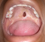
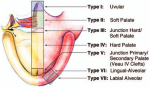
Incidence and location of fistulas: in the entire cohort, 22 patients developed a postoperative fistula (Figure 1), representing an overall incidence of 34.92% (Table 1). Regarding location, almost all fistulas (90.9%, n=20) were junctional (Figure 2), while 9.1% (n=2) were located in the soft palate. No pre-alveolar or alveolar fistulas were observed.
Risk factors associated with fistula: the Bivariate analysis (Table 2) revealed the following associations: defect diameter: a cleft diameter greater than 2 cm was significantly associated with an increased risk of fistula (OR=3.67; 95% CI: 1.00-13.34; p=0.027); laterality: bilateral clefts showed a trend toward a higher risk of fistula compared to unilateral clefts (OR=3.72; 95% CI: 0.79-17.40); although this difference did not reach statistical significance (p=0.079); sex: female sex was associated with a 2.2-fold increased risk of developing a fistula (95% CI: 0.77-6.798), but this association was not statistically significant (p=0.071); surgical technique: the Wardill technique was the most used (73.01). Although it showed a tendency towards a higher fistula rate (OR=1.5), the difference was not statistically significant (p=0.454) compared to the other techniques in this series.
This study reports a postoperative palatal fistula rate of 34.92% in Lubumbashi. This rate falls within the upper range of data reported in the international literature, which generally ranges from 5% to 35%, but it is higher than the averages of large series from high-income countries (often around 5% to 10%) [5,6]. This disparity may be explained by the late presentation of patients, the initial width of the clefts, and the technical constraints related to the context. The major finding of our study is the statistically significant correlation between defect width (>2 cm) and the occurrence of a fistula (p=0.027). This observation is universally recognized: a wide cleft requires more extensive dissection and generates increased tension on the suture lines, compromising the vascularization of the mucoperiosteal flaps and favoring necrosis or dehiscence [7-9].
We also noted a trend (not statistically significant, probably due to sample size) toward an increased risk of fistula in cases of bilateral clefts (OR=3.72) and in female patients. The anatomical complexity of bilateral clefts, often wider and with a free vomer, makes closure of the nasal layer particularly difficult, which corroborates the observations of other authors [10].
Regarding surgical techniques, the Wardill palatoplasty (push-back) was the most frequently performed in our series. Although this technique allows lengthening of the soft palate, it leaves areas of exposed bone laterally, which could theoretically increase the risk of fistula compared to tension-free closure techniques such as Furlow's double opposing Z-plasty or Bardach's two-flap palatoplasty [9,11]. However, our study did not show a statistically significant difference, which underlines that the surgeon's technical mastery and tension management are often more decisive than the technique itself.
Study limitations: the relatively small sample size (n=63) limits the statistical power to detect weak associations (such as the impact of sex or surgical technique) and precludes multivariate analysis. Additionally, the duration of postoperative follow-up was not uniformly extended, which could lead to an underestimation of late-presenting fistulas.
Perspectives: would the implementation, if possible, of pre-surgical orthopedic techniques (nasoalveolar molding) reduce the risk of fistula?
Postoperative palatal fistula is a frequent complication after cleft palate repair in our setting, occurring predominantly at the junction of the hard and soft palates. The initial cleft width (>2 cm) represents a statistically significant risk factor for fistula development in our series.
What is known about this topic
- Postoperative palatal fistula is a frequent complication following cleft palate repair, with incidence rates varying widely in the international literature;
- The initial width of the cleft, laterality, and the surgical technique are recognized as potential predictors of fistula formation;
- Data regarding the incidence and specific risk factors for this complication in sub-Saharan Africa, particularly in the Democratic Republic of Congo, remain scarce.
What this study adds
- This study reports a high incidence (34.92%) of postoperative palatal fistulas in Lubumbashi, predominantly located at the hard and soft palate junction.
- It identifies an initial cleft defect width greater than 2 cm as a statistically significant risk factor for fistula development in this population;
- The findings highlight the need for tension-free closure techniques and rigorous preoperative assessment to mitigate complications in resource-limited settings.
The authors declare no competing interests.
All the authors read and approved the final version of this manuscript.
Table 1: incidence and anatomical location of postoperative palatal fistulas following cleft palate repair in 63 patients treated across three healthcare facilities in Lubumbashi (University Clinics, Rwashi Military Hospital, and Afia/Don Bosco Polyclinic) over a 14-month period
Table 2: bivariate analysis of demographic, clinical, and surgical risk factors associated with postoperative palatal fistula after cleft palate repair in Lubumbashi, Democratic Republic of Congo (N=63, 14-month study period)
Figure 1: palatal fistula 1 month after palatoplasty according to Wardill
Figure 2: Pittsburgh classification of palatal fistulas
- Dao AM, Goudy SL. Cleft Palate Repair, Gingivoperiosteoplasty, and Alveolar Bone Grafting. Facial Plast Surg Clin North Am. 2016 Nov;24(4):467-476. PubMed | Google Scholar
- Cohen SR, Kalinowski J, LaRossa D, Randall P. Cleft palate fistulas: a multivariate statistical analysis of prevalence, etiology, and surgical management. Plast Reconstr Surg. 1991 Jun;87(6):1041-7. PubMed | Google Scholar
- Buller M, Jodeh D, Qamar F, Wright JM, Halsey JN, Rottgers SA. Cleft Palate Fistula: A Review. Eplasty. 2023 Feb 1;23:e7. eCollection 2023. PubMed | Google Scholar
- Mbuyi-Musanzayi S, Kayembe TJ, Kashal MK, Lukusa PT, Kalenga PM, Tshilombo FK et al. Non-syndromic cleft lip and/or cleft palate: Epidemiology and risk factors in Lubumbashi (DR Congo), a case-control study. J Craniomaxillofac Surg. 2018 Jul;46(7):1051-1058. PubMed | Google Scholar
- Hardwicke JT, Landini G, Richard BM. Fistula incidence after primary cleft palate repair: a systematic review of the literature. Plast Reconstr Surg. 2014 Oct;134(4):618e-627e. PubMed | Google Scholar
- Bykowski MR, Naran S, Winger DG, Losee JE. The Rate of Oronasal Fistula Following Primary Cleft Palate Surgery: A Meta-Analysis. Cleft Palate Craniofac J. 2015 Jul;52(4):e81-7. PubMed | Google Scholar
- Parwaz MA, Sharma RK, Parashar A, Nanda V, Biswas G, Makkar S. Width of cleft palate and postoperative palatal fistula--do they correlate? J Plast Reconstr Aesthet Surg. 2009 Dec;62(12):1559-63. PubMed | Google Scholar
- Basta MN, Silvestre J, Stransky C, Solot C, Cohen M, McDonald-McGinn D et al. A 35-year experience with syndromic cleft palate repair: operative outcomes and long-term speech function. Ann Plast Surg. 2014 Dec;73 Suppl 2:S130-5. PubMed | Google Scholar
- Jacob MF, Prearo GA, Brosco TV, Silva HL, Dutka JD. Fistula after primary palatoplasty: consensus among plastic surgery and Speech-Language Pathology. Revista Brasileira de Cirurgia Plástica. 2020;35:142-8. Google Scholar
- Deshpande GS, Campbell A, Jagtap R, Restrepo C, Dobie H, Keenan HT et al. Early Complications After Cleft Palate Repair: A Multivariate Statistical Analysis of 709 Patients. J Craniofac Surg. 2014 Sep;25(5):1614-8. PubMed | Google Scholar
- Horswell BB, Chou J. Does the Children's Hospital of Philadelphia Modification Improve the Fistula Rate in Furlow Double-Opposing Z-Plasty? J Oral Maxillofac Surg. 2020 Nov;78(11):2043-2053. PubMed | Google Scholar
Search
This article authors
On Pubmed
On Google Scholar
Citation [Download]
Navigate this article
Similar articles in
Key words
Tables and figures

Article metrics
Recently from the PAMJ




Authors´ services
