
Extranodal marginal zone lymphoma: epidemiological, clinical features and treatment outcomes
Manel Rebah, Malek Sayedi, Wafa Miled, Dorra Jaber, Raoudha Mansouri, Raihane Benlakhal, Karima Kacem
Corresponding author: Manel Rebah, Adult Clinical Haematology Department, Aziza Othmana Hospital, Tunis, Tunisia 
Received: 25 Aug 2025 - Accepted: 13 Aug 2026 - Published: 07 Oct 2026
Domain: Oncology
Keywords: Extranodal B cell lymphoma, clinical , treatment
Funding: This work received no specific grant from any funding agency in the public, commercial, or non-profit sectors.
©Manel Rebah et al. Pan African Medical Journal (ISSN: 1937-8688). This is an Open Access article distributed under the terms of the Creative Commons Attribution International 4.0 License (https://creativecommons.org/licenses/by/4.0/), which permits unrestricted use, distribution, and reproduction in any medium, provided the original work is properly cited.
Cite this article: Manel Rebah et al. Extranodal marginal zone lymphoma: epidemiological, clinical features and treatment outcomes. Pan African Medical Journal. 2026;55:65. [doi: 10.11604/pamj.2026.55.65.49140]
Available online at: https://www.panafrican-med-journal.com//content/article/55/65/full
 PDF in process
 Case study 

Extranodal marginal zone lymphoma: epidemiological, clinical features and treatment outcomes
Extranodal marginal zone lymphoma: epidemiological, clinical features and treatment outcomes
Manel Rebah1,&,  Malek Sayedi1, Wafa Miled1, Dorra Jaber1, Raoudha Mansouri1, Raihane Benlakhal1, Karima Kacem1
&Corresponding author
Extranodal marginal zone lymphoma (MZL) represents the most common form of MZL. Our study aimed to describe the epidemiological and clinical aspects and treatment outcomes of patients with extranodal MZL, as well as the factors associated with prognosis. This retrospective descriptive study included patients treated for untransformed extranodal MZL in the clinical haematology department of Aziza Othmana Hospital between January 2016 and December 2021. The classifications used were those of Ann Arbour and Ann Arbour modified by Mushoff for non-gastric and gastric localisations, respectively. Therapeutic evaluation was made according to the Cheson criteria for non-gastric localisations and GELA criteria. In our study, 35 patients were noted with a median age of 57 years (34-83). The sex ratio was 2.5. Thirteen gastric localisations were noted (37%). Non-gastric localisations were distributed as follows: 10 ocular, 7 pulmonary and 5 otorhinolaryngeal. Performance status was less than or equal to 1 in 85% of cases. Bone marrow involvement was noted in 24% of cases. Localised stage (I-II) was observed in 63% of cases. Regarding treatment: two patients had a watch-and-wait strategy, 9 had Helicobacter pylori eradication, one had immunotherapy, and 22 underwent immunochemotherapy. Evaluation after first-line therapy showed response in 19/28 (68%): 13 complete remissions, 6 partial responses. At the last count, global response was obtained in 23/28 (82%): 18 complete remissions,5 partial responses. Grade IV neutropenia was observed in 37% of cases. One patient relapsed. Four patients had died, two of them from the disease. In five years, global survival (GS) was 78%. Only osteomedullary infiltration significantly impacted GS (p=0,002). Extranodal MZL, with its varied clinical presentations, remains an indolent lymphoma with a good prognosis and satisfactory therapeutic response to immunochemotherapy.

Extranodal marginal zone lymphoma (MZL) is an indolent B-cell lymphoma, accounting for 7% of non-Hodgkin lymphomas (NHL) [1]. It can involve both lymphoid and non-lymphoid organs, with the extranodal form being the most common. Its etiopathogenic mechanism is driven by chronic inflammatory processes linked to persistent antigenic stimulation (e.g., microbial agents), immune-mediated mechanisms (e.g., autoantibodies) or additional molecular pathways. Clinical presentation varies depending on the involved site. Therapeutic management depends on both disease location and stage, though standardised guidelines remain lacking. In Tunisia, few studies have addressed this entity. A better characterisation of these lymphomas could optimise the management of extranodal MZL patients. Our study aimed to describe the epidemiological and clinical features, as well as treatment outcomes, in a series of extranodal MZL patients, and to identify potential prognostic factors.

Materials and methods
This was conducted as a retrospective descriptive study in the Clinical Haematology Department of Aziza Othmana Hospital between 2016 and 2021. Patients with an EMZL diagnosis according to World Health Organization (WHO) 2022 criteria were included [2]. The diagnosis was confirmed by a biopsy of the extranodal site on the basis of morphological and immunohistochemical (CD5, CD10, CD23) characteristics. Patients with transformed high-grade lymphoma were not included. We excluded patients with incomplete medical records. We recorded epidemiological characteristics of the patients (age, gender), disease presentation and first initial workup at initial evaluation: Blood count, lactate dehydrogenase, bone marrow biopsy (BMB) and imaging computed tomography (CT) or magnetic resonance imaging (MRI). We noted the treatment modalities and the evolution. The staging of the disease was based on the Ann Arbour classification for non-gastric extranodal MZL and Ann Arbour modified by Mushoff for gastric localisations. Response criteria were based on Cheson and GELA criteria, respectively, for gastric and non-gastric localisations [3,4]. Statistical data was analyzed by Spss 25 version. Survival analyses were performed via the Kaplan-Meier method. Prognostic factors were analysed through univariate analysis (Statistical significance was set at p < 0.05).
Results
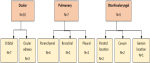
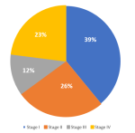
We recorded 35 cases of extranodal MZL patients between 2016 and 2021. Epidemiological, clinical and biological features were summarised in Table 1. For gastric localisations, there were five antral locations, four fundic, one in the gastric body, one antrofundic and two pangastric. Non-gastric localisations were detailed in Figure 1. Localised stage (I-II) was noted in 63% of cases, and extended stage (III-IV) was observed in 37% (Figure 2). As for the initial therapy setting, nine patients with gastric MZL had Helicobacter pylori (HP) eradication, one had immunotherapy, and 22 underwent immunochemotherapy (8 R-CHOP, 12 R-CLB, 2 R-CVP). One patient died before initiation of therapy, and two had a watch-and-wait strategy for ocular localisation.
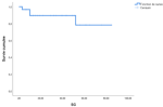
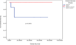
Among the 32 patients treated in the initial setting, 28 were evaluable. Sixty eight percent were showing response (46% complete response (CR)). Six patients had second-line treatment which four obtained CR. Two patients were refractory to treatment which one was salvaged by third-line therapy and an autologous stem cell transplantation and obtained a CR. During follow-up, one patient had a relapse after 8 months of CR and was awaiting salvage therapy, and one patient progressed after three months of partial response. The five-year overall survival (OS) rate was 78% (Figure 3). Four patients died which two of disease progression. The analysis of factors associated with modified OS showed that bone marrow infiltration was an important prognostic factor for survival (p=0,002), as shown in Figure 4.
Marginal zone lymphoma (MZL) is a rare indolent B-cell malignancy accounting for 7% of non-Hodgkin lymphomas in Western countries [5]. A United States study reported that extranodal MZL represented 61% of 7,460 MZL cases, with annual incidence increasing by 1%. A multinational study across 24 countries showed higher extranodal MZL frequency in developed countries (8.8%) versus developing nations (5.2%) [6]. Peak incidence occurs between 50-60 years (median 57 years in our cohort, range 34-83). According to various studies, there is no significant gender predominance, except for certain parotid (p=0.03) and breast localisations [7]. In our study, the male-to-female ratio was 2.5.
Risk factors: extranodal MZL is associated with chronic bacterial infections. The most proven association is with HP with gastric localisation [8]. Other emerging associations exist, such as Borrelia burgdorferi (cutaneous), Chlamydophila psittaci (ocular), Achromobacter xylosoxidans (pulmonary) [9]. Other viral infection associations were identified, such as Human immunodeficiency virus (HIV) and Hepatitis C virus [10]. In our study, we noted 7 cases of HP infection and one case of HIV in cavum localisation. Other chronic immune stimulations may present in autoimmune disorders (Hashimoto thyroiditis, Sjögren syndrome) [11]. Different translocations were identified through different localisations and have the potential to transform into high-grade lymphoma. The most frequent one is the translocation and is associated with HP-negative gastric and pulmonary localisations [12,13].
The most frequent localisation is the stomach, which represents 30 to 35% [14,15] (Table 2). It may present with nausea, vomiting, dyspepsia or rarely B symptoms [16]. Histopathology may show dense lymphoid infiltrate associated with plasmacytoid differentiation or lymphoepithelial lesions. There is no specific marker for extranodal MZL [17]. For initial workup, CT remains the gold standard for imaging in this type of lymphoma. MRI may be essential in salivary glands and ocular localisations [18]. Positron emission tomography is not obligatory, but it can detect bulky masses, lymph node dissemination, bone marrow involvement and suspicion of transformation. Endoscopy can assess mucosal changes for gastric locations (erosions, ulcers, wall thickening) [19], but endoscopic ultrasound can better evaluate wall infiltration and regional nodes [20]. Treatment approaches may differ depending on the stage and localisation. Eradication of HP is recommended in gastric localisations and consists of an association of a proton pump inhibitor with 2 or 3 antibiotics. Assessment of eradication can be done after 6 weeks of treatment, and evaluation of lymphoma after 3 months by a biopsy. Radiotherapy remains the preferred treatment option for localised stage [21]. Surgery is rarely indicated, especially in gastric localisations, because of its association with comorbidities [22]. For systemic disease, immunotherapy and/or chemotherapy may be prescribed.
The standard treatment for this type of lymphoma is the rituximab-chlorambucil (R-Clb) association, which was approved by the IELSG19 study [23]. R-CHOP may be indicated in some aggressive forms [24]. The use of rituximab-bendamustine (R-benda) has also proven its efficacy, with a need for viral prophylaxis [25,26]. Our study allowed a study of a malignant lymphoid hemopathy which had heterogenous clinic characteristics and various treatment approaches. But it had some limitations due to its retrospective character and the insufficient number of patients.
Extranodal MZL is an indolent lymphoid hemopathy that often remains localised for extended periods. The most common site is gastric, where H. pylori eradication therapy has revolutionised management. Radiotherapy may be considered for localised disease, while immunochemotherapy remains the cornerstone for advanced stages, with R-Clb as the standard regimen and R-CHOP/RCVP reserved for aggressive forms. R-Benda can be offered as second-line therapy. Bruton's tyrosine kinase inhibitors have emerged for refractory or relapsed cases. However, improved understanding of ethiopathogenic mechanisms may allow discovery of more effective and less toxic therapeutic targets.
The authors declare no competing interests.
Manel Rebah: conception and design of the study and drafting of the manuscript. Manel Rebah, Malek Sayedi, Wafa Miled, Dorra Jaber and Raoudha Mansouri: data collection. Manel Rebah and Malek Sayedi: data analysis and interpretation. Manel Rebah, Malek Sayedi, Wafa Miled, Dorra Jaber, Raoudha Mansouri, Karima Kacem and Raihane Benlakhal: critical revision of the manuscript. Karima Kacem and Raihane Benlakhal: supervision of the study. All authors read and approved the final version of the manuscript.
Table 1: epidemiological, clinical and biological characteristics of patients
Table 2: sites distribution according to various studies
Figure 1: non-gastric localisations' distribution
Figure 2: distribution of stages in EMZL
Figure 3: global survival curve
Figure 4: global survival according to osteomedullary infiltration
- Cerhan JR, Habermann TM. Epidemiology of marginal zone lymphoma. Ann Lymphoma. 2021 Mar ;5:1-38. PubMed | Google Scholar
- Campo E, Jaffe ES, Cook JR, Quintanilla-Martinez L, Swerdlow SH, Anderson KC et al. The international consensus classification of mature lymphoid neoplasms: a report from the clinical advisory committee. Blood. 2022 Sep;140(11):1229-53. PubMed | Google Scholar
- Cheson BD, Pfistner B, Juweid ME, Gascoyne RD, Specht L, Horning SJ et al. Revised response criteria for malignant lymphoma. J Clin Oncol. 2007 Feb;25(5):579-86. PubMed | Google Scholar
- Copie-Bergman C, Wotherspoon AC, Capella C, Motta T, Pedrinis E, Pileri SA et al. Gela histological scoring system for post-treatment biopsies of patients with gastric MALT lymphoma is feasible and reliable in routine practice. Br J Haematol. 2013 Jan;160(1):47-52. PubMed | Google Scholar
- Bertoni F, Coiffier B, Salles G, Stathis A, Traverse-Glehen A, Thieblemont C et al. MALT lymphomas: pathogenesis can drive treatment. Oncology. 2011 Nov;25(12):1134-42. PubMed | Google Scholar
- Perry AM, Diebold J, Nathwani BN, MacLennan KA, Müller-Hermelink HK, Bast M et al. Non-hodgkin lymphoma in the developing world: review of 4539 cases from the international non-hodgkin lymphoma classification project. Haematologica. 2016 Oct;101(10):1244-50. PubMed | Google Scholar
- Kiesewetter B, Lukas J, Dolak W, Simonitsch-Klupp I, Mayerhoefer ME, Raderer M. Gender aspects in extranodal marginal zone b-cell lymphoma of the mucosa-associated lymphoid tissue: does sex matter? Oncology. 2016 Aug;91(5):243-50. PubMed | Google Scholar
- Ferreri AJ, Govi S, Ponzoni M. Marginal zone lymphomas and infectious agents. Semin Cancer Biol. 2013 Dec;23(6):431-40. PubMed | Google Scholar
- Zucca E, Bertoni F, Vannata B, Cavalli F. Emerging role of infectious etiologies in the pathogenesis of marginal zone b-cell lymphomas. Clin Cancer Res. 2014 Oct;20(20):5207-16. PubMed | Google Scholar
- Arcaini L, Burcheri S, Rossi A, Paulli M, Bruno R, Passamonti F et al. Prevalence of HCV infection in nongastric marginal zone B-cell lymphoma of MALT. Ann Oncol. 2007 Feb;18(2):346-50. PubMed | Google Scholar
- Thieblemont C, Bertoni F, Copie-Bergman C, Ferreri AJ, Ponzoni M. Chronic inflammation and extra-nodal marginal-zone lymphomas of MALT-type. Semin Cancer Biol. 2014 Feb;24:33-42. PubMed | Google Scholar
- Troppan K, Wenzl K, Neumeister P, Deutsch A. Molecular pathogenesis of MALT lymphoma. Gastroenterol Res Pract. 2015 Nov;2015:1-10. PubMed | Google Scholar
- Ronchi A, Montella M, Panarese I, Costanzo RM, Aquino G, De Chiara A, Franco R, Marino FZ. MALT gastric lymphoma: An update of pathogenetic features. WCRJ. 2016 Jan 1;3(2):1-0. Google Scholar
- Thieblemont C, Coiffier B. Management of marginal zone lymphomas. Curr Treat Options Oncol. 2006 May;7(3):213-22. PubMed | Google Scholar
- Jeong SH, Hyun SY, Choi JS, Kim HM. Trends of incidence and survival rates of mucosa-associated lymphoid tissue lymphoma in the Korean population: analysis of the Korea central cancer registry database. J Korean Med Sci. 2020 Sep;35 (36):1-10. PubMed | Google Scholar
- Zinzani PL. The many faces of marginal zone lymphoma. Hematology Am Soc Hematol Educ Program. 2012 Dec;2012:426-32. PubMed | Google Scholar
- Vuka K, Dimov D, Katic K. The histopathology and immunohistology of gastric MALT lymphoma. Arch Oncol. 2004 Jan;12:5-6. Google Scholar
- Zucca E, Arcaini L, Buske C, Johnson PW, Ponzoni M, Raderer M et al. Marginal zone lymphomas: ESMO Clinical Practice Guidelines for diagnosis, treatment and follow-up .Ann Onco. 2020 Jan;31(1):17-29. PubMed | Google Scholar
- Tsai HF, Hsu PN. Interplay between Helicobacter pylori and immune cells in immune pathogenesis of gastric inflammation and mucosal pathology. Cell Mol Immunol. 2010 Jul;7(4):255-9. PubMed | Google Scholar
- Warrick J, Luo J, Robirds D, Branson J, Frater JL, Kreisel F et al. Gastrointestinal lymphomas in a North American population: clinicopathologic features from one major central-midwestern United States tertiary care medical center. Diagn Pathol. 2012 Jun;7:1-9. PubMed | Google Scholar
- Chey WD, Leontiadis GI, Howden CW, Moss SF. ACG clinical guideline: treatment of helicobacter pylori infection. Am J Gastroenterol. 2017 Feb;112(2):212-39. PubMed | Google Scholar
- Alderuccio JP, Kahl BS. Current Treatments in Marginal Zone Lymphoma. Oncology (Williston Park). 2022 Apr 6;36(4):206-215. PubMed | Google Scholar
- Hammel P, Haioun C, Chaumette MT, Gaulard P, Divine M, Reyes F et al. Efficacy of single-agent chemotherapy in low-grade B-cell mucosa-associated lymphoid tissue lymphoma with prominent gastric expression. J Clin Oncol. 1995 Oct;13(10):2524?9. PubMed | Google Scholar
- Thieblemont C, Zucca E. Clinical aspects and therapy of gastrointestinal MALT lymphoma. Best Pract Res Clin Haematol. 2017 Mar;30(1-2):109-17. PubMed | Google Scholar
- Alderuccio JP, Arcaini L, Watkins MP, Beaven AW, Shouse G, Epperla N et al. An international analysis evaluating frontline bendamustine with rituximab in extranodal marginal zone lymphoma. Blood Adv. 2022 Apr;6(7):2035-44. PubMed | Google Scholar
- Morigi A, Argnani L, Lolli G, Broccoli A, Pellegrini C, Nanni L et al. Bendamustine-rituximab regimen in untreated indolent marginal zone lymphoma: experience on 65 patients. Hematol Oncol. 2020 Oct;38(4):487-92. PubMed | Google Scholar
Search
This article authors
On Pubmed
On Google Scholar
Citation [Download]
Navigate this article
Similar articles in
Key words
Tables and figures

Article metrics
Recently from the PAMJ





Authors´ services
