Comparative analysis of IL-1β and IFN-γ expression after subcutaneous implantation of DBBM, FDBB, and dc-FDBB scaffolds in a Rattus norvegicus rat model
Ardian Pradana, Sekti Anggara, David Buntoro Kamadjaja, Aries Muharram
Corresponding author: Ardian Pradana, Department of Oral and Maxillofacial Surgery, Faculty of Dental Medicine, Universitas Airlangga, Surabaya, Indonesia 
Received: 14 Apr 2026 - Accepted: 11 Oct 2026 - Published: 11 Nov 2026
Domain: Molecular Biology
Keywords: Xenograft, bovine bone scaffold, IL-1β, IFN-γ, immune response
Funding: This work received no specific grant from any funding agency in the public, commercial, or non-profit sectors.
©Ardian Pradana et al. Pan African Medical Journal (ISSN: 1937-8688). This is an Open Access article distributed under the terms of the Creative Commons Attribution International 4.0 License (https://creativecommons.org/licenses/by/4.0/), which permits unrestricted use, distribution, and reproduction in any medium, provided the original work is properly cited.
Cite this article: Ardian Pradana et al. Comparative analysis of IL-1β and IFN-γ expression after subcutaneous implantation of DBBM, FDBB, and dc-FDBB scaffolds in a Rattus norvegicus rat model. Pan African Medical Journal. 2026;55:116. [doi: 10.11604/pamj.2026.55.116.52748]
Available online at: https://www.panafrican-med-journal.com//content/article/55/116/full
 PDF in process
Research 

Comparative analysis of IL-1β and IFN-γ expression after subcutaneous implantation of DBBM, FDBB, and dc-FDBB scaffolds in a Rattus norvegicus rat model
Comparative analysis of IL-1β and IFN-γ expression after subcutaneous implantation of DBBM, FDBB, and dc-FDBB scaffolds in a Rattus norvegicus rat model
Ardian Pradana1,&, Sekti Anggara1, David Buntoro Kamadjaja1, Aries Muharram1
&Corresponding author
Introduction: Bovine xenograft scaffolds are bone grafts derived from bovine bone, making them abundantly available. Deproteinized bovine bone mineral (DBBM), freeze-dried bovine bone (FDBB), and freeze dried bovine bone (dc-FDBB) are commonly used scaffold types. IL-1β and IFN-γ are proinflammatory cytokines that play an important role in bone graft success, as an inflammatory response is needed to clear necrotic tissue but must not be excessive and interfere with osteointegration. This study aimed to compare the expression of IL-1β and IFN-γ following subcutaneous implantation of the bovine scaffolds DBBM, FDBB, and dc-FDBB on days 2, 7, and 14 in white rats (Rattus norvegicus).
Methods: this experimental study used 60 Rattus norvegicus rats divided into four groups: sham operation (SO) (control), DBBM, FDBB, and dc-FDBB. We observed each group on days 2, 7, and 14, with five samples at each time point. The expression of IL-1β and IFN-γ was evaluated using immunohistochemical staining, followed by manual cell counting and analysis using a one-way ANOVA test.
Results: IL-1β expression in the FDBB, DBBM, and dc-FDBB groups showed no significant differences on days 2, 7, and 14, but there were significant differences between the treatment groups and the control group. Similarly, IFN-γ expression showed no significant differences among FDBB, DBBM, and dc-FDBB at all observation time points. On day 2, there was no significant difference between the treatment and control groups, but on days 7 and 14, significant differences were observed between them.
Conclusion: subcutaneous implantation of FDBB, DBBM, and dc-FDBB scaffolds on days 2, 7, and 14 resulted in IL-1β and IFN-γ expression that showed descriptive variation but no statistically significant differences among the scaffold groups. These findings indicate that all three types of xenografts elicit a low and comparable immune response, supporting their suitability as bone reconstruction materials from an immunological biocompatibility perspective.

Bone is a dynamic, highly vascularized tissue with lifelong regenerative capacity. This natural process enables repair following minor injury; however, when damage exceeds the critical size defect (CSD), spontaneous healing is no longer possible, and clinical intervention is required. Large bone defects may result from trauma, developmental abnormalities, tumor resection, or infection, and remain a major clinical challenge in medicine and maxillofacial surgery. Epidemiological data indicate that between 2010 and 2025, the incidence of bone fractures in Europe increased by up to 28% annually, accompanied by an approximately 25% rise in economic burden, thereby driving intensified research in bone regenerative therapies [1].
One of the primary strategies in bone tissue engineering is the use of bone grafts to replace or stimulate new bone formation. Common types include autografts, allografts, and xenografts. Autologous bone grafts remain the gold standard due to their excellent osteoinductive, osteoconductive, and osseointegrative properties, enabling the formation of strong and well-organized bone tissue. However, their use is limited by donor site pain and morbidity, restricted tissue availability, risk of blood loss, and longer operative time [2].
As alternatives, numerous studies have explored allografts, xenografts, and alloplasts to overcome these limitations. Ideally, bone graft substitutes should exhibit biomechanical stability, osteoconductive and osteoinductive properties, controlled degradation matching new bone formation, and the ability to support angiogenesis and osteogenic cell migration. Nevertheless, allografts still face challenges such as suboptimal osseointegration, immune reactions, and potential pathogen transmission, which has led to increasing interest in xenografts due to their high availability, lower cost, and scalability for mass production [3,4].
Xenografts are derived from different species, commonly bovine bone, and are generally osteoconductive. With appropriate processing, they can achieve high biocompatibility and reduced immunogenicity [5]. One key strategy to minimize immunogenicity is decellularization, which removes cellular components while preserving the extracellular matrix (ECM) structure and composition. Decellularized freeze-dried bovine bone represents an advancement of freeze-dried bovine bone, where decellularization eliminates residual DNA and antigens such as the Gal epitope that may trigger immune responses [2].
Three main types of bovine-derived bone scaffolds are commonly used: deproteinized bovine bone mineral, freeze-dried bovine bone xenograft, and decellularized freeze-dried bovine bone. Deproteinized bovine bone mineral is produced through deproteinization, resulting in high biocompatibility but lacking organic components, thus exhibiting no osteoinductive properties. In contrast, FDBB retains organic components such as growth factors beneficial for osteogenesis, but carries a risk of immune response due to residual cellular materials. Decellularized freeze-dried bovine bone was developed to address these limitations by preserving the natural ECM structure while reducing immunogenicity, thereby enhancing its potential for optimal bone regeneration [1].
The interaction between scaffold ECM and the innate immune system is influenced by damage-associated molecular patterns (DAMPs) released during tissue injury or graft processing. Damage-associated molecular patterns such as DNA, reactive oxygen species (ROS), hyaluronic acid, and fibronectin fragments are recognized by pattern recognition receptors (PRRs) on immune cells. Pattern recognition receptor activation triggers proinflammatory gene expression and the secretion of cytokines such as interleukin-1 beta (IL-1β), interferon-gamma (IFN-γ), and TNF-α, which play roles in immune cell recruitment and macrophage polarization toward the M1 phenotype [6].
In bone regeneration, IL-1β and IFN-γ are critical in regulating the balance between inflammation and osseointegration. IL-1β contributes to the early inflammatory phase and supports osteogenesis and scaffold remodeling at physiological levels, but may induce bone resorption when excessively expressed. Meanwhile, IFN-γ is involved in cellular immunity through Th1 cell differentiation and macrophage activity regulation [7]. Therefore, IL-1β and IFN-γ play crucial roles in the success of bone graft implantation. An adequate inflammatory response is necessary to clear necrotic tissue, but excessive inflammation may impair osseointegration. Thus, further research is needed to evaluate the expression of IL-1β and IFN-γ following subcutaneous implantation of bovine bone scaffolds, including DBBM, FDBB, and dc-FDBB.

Study design: this study was an in vivo experimental laboratory study conducted to analyze the expression of interleukin-1 beta (IL-1β) and interferon-gamma (IFN-γ) in paraffin block specimens following subcutaneous implantation of bovine bone scaffolds, including deproteinized bovine bone mineral, freeze-dried bovine bone, and decellularized freeze-dried bovine bone. The study employed a post-test-only control group design to compare cytokine expression among treatment and control groups.
Experimental groups and design: the experimental animals were divided into four groups: DBBM, FDBB, dc-FDBB, and a control group (same operation, SO). The treatment groups consisted of Rattus norvegicus implanted with the respective scaffolds, whereas the control group underwent identical surgical procedures without scaffold implantation. Observations were conducted at three time points, namely day 2, day 7, and day 14 post-implantation, to evaluate temporal changes in inflammatory and immune responses.
Sample size determination: the minimum sample size was estimated using the single-proportion formula described by Lwanga and Lemeshow (8]:
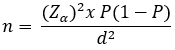
In this formula, n represents the minimum required sample size; Zα is the standard normal deviate corresponding to the selected confidence level (1.96 for a 95% confidence level); P is the anticipated proportion of the outcome in the population; (1 - P) is the complementary proportion; and d is the desired absolute precision or margin of error. The original study protocol reported a minimum requirement of three animals per group. A 10% allowance for potential sample loss was considered, giving a minimum of four animals per group after rounding up. To provide an additional margin and maintain balanced allocation across the experimental groups and observation time points, we included five animals in each group at each time point. The study therefore included 60 animals in total (4 groups x 3 observation time points x 5 animals). IL-1β and IFN-γ were assessed in each specimen, resulting in 120 cytokine measurements; these represent two outcome measurements per specimen, not 120 independent animals.
Variables: the independent variables in this study were the types of bovine bone scaffolds (DBBM, FDBB, and dc-FDBB). The dependent variables were the expression levels of IL-1β and IFN-γ identified through immunohistochemical staining. Controlled variables included the animal model (Wistar strain Rattus norvegicus), scaffold size and shape, surgical procedures, environmental conditions, and animal care protocols to ensure consistency throughout the experiment.
Materials and scaffold preparation: the control group, referred to as the same operation (SO) group, underwent anesthesia, incision, pocket formation, and suturing without implantation of any scaffold, allowing differentiation between surgical trauma and biomaterial-induced responses. The DBBM scaffold was derived from bovine cancellous femoral bone processed through high-temperature deproteinization (approximately 1000°C), resulting in an inorganic mineral matrix. The FDBB scaffold was produced by washing bovine cancellous bone with 3% hydrogen peroxide, followed by freeze-drying at -80°C and lyophilization to achieve moisture content below 10%, preserving organic components such as growth factors. The dc-FDBB scaffold was further processed by decellularization using 0.5% sodium dodecyl sulfate (SDS) to remove cellular components while maintaining extracellular matrix integrity. All scaffolds were standardized to dimensions of 5 x 5 x 3 mm, sterilized using gamma irradiation, and produced by the National Research and Innovation Agency (BRIN), Jakarta, Indonesia.
Histological preparation: paraffin block specimens obtained from subcutaneous tissue were sectioned at a thickness of 4 μm using a microtome. The sections were mounted on glass slides and subjected to deparaffinization with xylene, followed by rehydration using graded alcohol solutions (98%, 80%, and 50%) and rinsing with distilled water. Endogenous peroxidase activity was blocked by incubation with 3% hydrogen peroxide in methanol for 30 minutes at room temperature, followed by incubation with 3% normal horse serum for 20 minutes to reduce nonspecific binding. Each step was followed by washing with phosphate-buffered saline (PBS, pH 7.4). Antigen retrieval was performed using citrate buffer and microwave heating (5 minutes at high power and 10 minutes at low power), followed by cooling at room temperature and PBS washing.
Immunohistochemical analysis: immunohistochemical staining was performed to detect IL-1β and IFN-γ expression. The specimens were incubated overnight with primary antibodies at a dilution of 1:200. After staining, positive cells were identified by brown chromogen deposition in the cytoplasm. Quantitative analysis was conducted by manually counting positively stained cells in 20 fields of view under a light microscope at 1000x magnification. IL-1β-positive cells were typically observed in inflammatory cells such as macrophages and mononuclear cells, while IFN-γ-positive cells appeared as small to medium-sized cells with brown cytoplasmic staining.
Data analysis: the data obtained were quantitative and analyzed using statistical methods. Descriptive analysis was first performed, followed by testing for normality using the Shapiro-Wilk test, where a p-value greater than 0.05 indicated normal distribution. Homogeneity of variance was assessed using Levene's test. If the data met the assumptions of normality and homogeneity, differences between groups were analyzed using one-way analysis of variance (ANOVA), followed by post hoc tests to determine pairwise differences. Statistical significance was determined at a predefined confidence level.Ethical approval and study location: the ethical approval for this study was obtained from the Health Research Ethical Clearance Commission, Faculty of Dental Medicine, Universitas Airlangga. The study was approved under certificate number 0701/HRECC.FODM/VII/2025. All experimental procedures were conducted in accordance with institutional ethical standards and guidelines for the use of laboratory animals.
This study was conducted using 60 Rattus norvegicus, which were divided into four groups: same operation (SO), DBBM scaffold, FDBB scaffold, and dc-FDBB scaffold groups. Each group consisted of five samples observed at three time points, namely day 2, day 7, and day 14. Throughout the experimental period, no mortality was observed among the animals. Sample termination and collection were performed at each designated time point according to the established protocol. All samples were found to be in good condition, embedded within subcutaneous tissue, and no scaffold exposure was observed in any group. The collected samples were processed into paraffin blocks, followed by immunohistochemical (IHC) staining using monoclonal antibodies against IL-1β and IFN-γ. The stained sections were then observed under a light microscope and quantitatively analyzed.
IL-1β expression: the evaluation of IL-1β expression using immunohistochemical staining demonstrated that on day 2, the highest mean expression was observed in the SO group (8.2 ± 1.30), followed by dc-FDBB (5.8 ± 1.48), FDBB (5.6 ± 1.14), and DBBM (5.2 ± 0.83). On day 7, IL-1β expression decreased in all groups, with the SO group remaining the highest (7.0 ± 1.58), followed by dc-FDBB and DBBM, which showed equal values (4.0 ± 1.58), and FDBB with the lowest expression (3.4 ± 1.14). By day 14, the decreasing trend continued, with the SO group showing a mean of 6.0 ± 1.58, dc-FDBB at 3.4 ± 1.51, FDBB at 3.0 ± 1.58, and DBBM at 2.8 ± 1.30 (Table 1).
Statistical analysis was performed to evaluate data distribution and group differences. Normality testing using the Shapiro-Wilk test showed that all groups at all time points had p-values greater than 0.05, indicating that the data were normally distributed. Homogeneity testing using Levene's test also demonstrated p-values greater than 0.05 (day 2: p = 0.744; day 7: p = 0.888; day 14: p = 0.986), confirming that the data were homogeneous across groups. Subsequently, one-way ANOVA analysis revealed statistically significant differences in IL-1β expression among groups at all time points, with p = 0.005 on day 2, p = 0.006 on day 7, and p = 0.013 on day 14. These findings indicate that IL-1β expression differed significantly among the experimental groups (Table 2). Post hoc analysis using Tukey HSD demonstrated that the significant differences consistently occurred between the SO group and all scaffold groups (DBBM, FDBB, and dc-FDBB) at each time point. On day 2, significant differences were observed between SO and DBBM (p = 0.006), SO and FDBB (p = 0.018), and SO and dc-FDBB (p = 0.030) (Table 3).
On day 7, post hoc multiple comparison analysis using the Tukey HSD test revealed p-values of 0.026 for the SO vs. DBBM group, 0.007 for the SO vs. FDBB group, and 0.026 for the SO vs. dc-FDBB group. For comparisons between FDBB and DBBM, the p-value was 0.918, and between FDBB and dc-FDBB, the p-value was also 0.918. Meanwhile, the comparison between dc-FDBB and DBBM showed a p-value of 1.000. These results indicate that comparisons between the SO group and all scaffold groups (FDBB, DBBM, and dc-FDBB) had p-values < 0.05, demonstrating statistically significant differences on day 7. In contrast, comparisons among DBBM, FDBB, and dc-FDBB groups showed p-values > 0.05, indicating no statistically significant differences among these scaffold groups (Table 4).
On day 14, the p-values were 0.018 for the SO vs. DBBM group, 0.028 for the SO vs. FDBB group, and 0.043 for the SO vs. dc-FDBB group. For comparisons between FDBB and DBBM, the p-value was 0.997, and between FDBB and dc-FDBB, the p-value was 0.974. Meanwhile, the comparison between dc-FDBB and DBBM showed a p-value of 0.920. These results indicate that comparisons between the SO group and all scaffold groups (DBBM, FDBB, and dc-FDBB) had p-values < 0.05, demonstrating statistically significant differences on day 14. In contrast, comparisons among DBBM, FDBB, and dc-FDBB groups showed p-values > 0.05, indicating no statistically significant differences.
IFN-γ expression: the evaluation of IFN-γ expression using immunohistochemical staining showed that on day 2, the SO, DBBM, and dc-FDBB groups had similar mean expression values (7.0 ± 1.581), while FDBB showed a slightly lower value (6.4 ± 1.14). On day 7, the highest expression was observed in the SO group (9.0 ± 1.581), followed by dc-FDBB (5.2 ± 0.836), FDBB (4.4 ± 1.14), and DBBM (4.0 ± 1.0). On day 14, the same pattern persisted, with the SO group showing the highest expression (7.8 ± 2.387), followed by dc-FDBB (4.6 ± 1.14), FDBB (3.2 ± 1.302), and DBBM (2.4 ± 1.14) (Table 5). Normality testing using the Shapiro–Wilk test indicated that all groups had p-values greater than 0.05, confirming normal distribution of data. Homogeneity testing using Levene's test also showed p-values greater than 0.05 (day 2: p = 0.88; day 7: p = 0.52; day 14: p > 0.05), indicating homogeneous variance across groups. One-way ANOVA analysis demonstrated that there was no statistically significant difference in IFN-γ expression on day 2 (p = 0.892). However, significant differences were observed on day 7 and day 14 (p < 0.001), indicating that group differences became more evident during later stages of observation (Table 6).
Subsequently, a post hoc test using the Tukey HSD test was performed to determine which groups showed significant differences. On day 2, no significant differences were found among the groups; therefore, post hoc analysis was not required. On day 7, the Tukey HSD test revealed p-values of less than 0.001 for the SO vs. FDBB and SO vs. DBBM groups, and 0.001 for the SO vs. dc-FDBB group. For comparisons between DBBM and FDBB, the p-value was 0.948, and between DBBM and dc-FDBB, the p-value was 0.397. Meanwhile, the comparison between dc-FDBB and FDBB showed a p-value of 0.707. These results indicate that comparisons between the SO group and all scaffold groups had p-values < 0.05, demonstrating statistically significant differences on day 7. In contrast, comparisons among the DBBM, FDBB, and dc-FDBB groups showed p-values > 0.05, indicating no statistically significant differences (Table 7).
On day 14, the p-values were less than 0.001 for the SO vs. DBBM group, 0.002 for the SO vs. FDBB group, and 0.026 for the SO vs. dc-FDBB group. For comparisons between FDBB and DBBM, the p-value was 0.853, and between FDBB and dc-FDBB, the p-value was 0.517. Meanwhile, the comparison between dc-FDBB and DBBM showed a p-value of 0.165. These results indicate that comparisons between the SO group and all scaffold groups had p-values < 0.05, demonstrating statistically significant differences. In contrast, comparisons among the dc-FDBB, DBBM, and FDBB groups showed p-values > 0.05, indicating no statistically significant differences on day 14.
Among the scaffold groups, dc-FDBB showed the highest IFN-γ expression compared to DBBM and FDBB after day 2, with values decreasing from 7.0 on day 2 to 5.2 on day 7 and 4.6 on day 14. The FDBB group demonstrated a steady decline from 6.4 on day 2 to 4.4 on day 7 and 3.2 on day 14. Similarly, the DBBM group showed a continuous decrease, with values of 7.0 on day 2, 4.0 on day 7, and 2.4 on day 14, representing the lowest expression among all scaffold groups.
Overall, all scaffold groups (DBBM, FDBB, and dc-FDBB) demonstrated a consistent reduction in IFN-γ expression over time, indicating a decrease in immune activation. In contrast, the SO group maintained higher IFN-γ levels throughout the observation period, suggesting a more sustained inflammatory and immune response in the absence of scaffold implantation. These findings indicate that scaffold implantation may help modulate and reduce IFN-γ-mediated immune responses.
The success of biomaterial application is largely influenced by the inflammatory response that occurs following implantation. Initial protein adsorption on the biomaterial surface triggers activation of both innate and adaptive immunity [9]. Macrophages are considered the primary effectors in the foreign body reaction (FBR). Direct interaction between macrophages and the biomaterial surface induces the secretion of proinflammatory cytokines such as IL-1β, TNF-α, and IL-6. These cytokines stimulate stromal cells and osteoblasts surrounding the graft to express Receptor Activator of Nuclear Factor kappa-B Ligand (RANKL) and suppress Osteoprotegerin (OPG), thereby enhancing osteoclastogenesis. In later phases, activated T cells, particularly Th1 and Th17, also express RANKL and TNF-α, further promoting osteoclast precursor differentiation. In contrast, Th2 and Treg cells, along with M2 macrophages, suppress inflammation and support osteogenesis through increased OPG and anti-inflammatory cytokines [9,10].
Prolonged inflammation leads to macrophage fusion into foreign body giant cells (FBGCs) in an attempt to phagocytose the implanted biomaterial, which subsequently promotes fibroblast recruitment, fibrous capsule formation, isolation, and potential premature scaffold failure. In addition to IL-1β, IFN-γ plays a central role in cell-mediated immunity, macrophage activation, and antigen presentation, all of which are critical in the host response to implanted scaffolds. In this study, IL-1β and IFN-γ expression were evaluated using immunohistochemical staining with monoclonal antibodies. The mean expression values were analyzed to represent cytokine activity in both innate and adaptive immune responses that may influence xenograft rejection [10,11].
Our study results demonstrated that IL-1β expression was highest on day 2, likely due to the presence of xenogeneic DNA within the scaffold material, which triggers an early inflammatory response. Neutrophils infiltrate the biomaterial within 24-48 hours and release enzymes and peptides that regulate further cell recruitment and degradation processes. In response to inflammatory stimuli, neutrophils undergo a unique cell death process known as NETosis, releasing neutrophil extracellular traps (NETs) containing serine proteases and antimicrobial peptides [12]. These NETs are recognized by macrophages as damage-associated molecular patterns (DAMPs), activating inflammatory signaling and promoting IL-1β production. Moreover, IL-1β produced by macrophages further enhances NET formation in neutrophils. By days 7 and 14, corresponding to the late inflammatory or early proliferative phase, IL-1β expression decreased, while growth factors and osteogenic markers began to increase. This reduction aligns with macrophage phenotype switching and decreased inflammatory signaling around the scaffold [13,14].
On day 7, IL-1β expression decreased across all groups, although the control group (SO) remained the highest (7.0 ± 1.58). Both dc-FDBB and DBBM showed similar values (4.0 ± 1.58), while FDBB was slightly lower (3.4 ± 1.14). This decrease reflects the transition from acute inflammation to tissue healing and integration, where well-decellularized graft materials exhibit lower inflammatory activity [15]. By day 14, the decreasing trend continued, with SO at 6.0 ± 1.58, dc-FDBB at 3.4 ± 1.51, FDBB at 3.0 ± 1.58, and DBBM at 2.8 ± 1.30. Persistent IL-1β expression in the SO group indicates unresolved inflammation, whereas lower levels in DBBM and FDBB suggest better control of chronic inflammation and improved integration potential [15].
Overall, IL-1β expression showed a consistent decline over time in all groups, with the SO group consistently exhibiting the highest levels. On day 2, IL-1β expression was highest in the SO group (8.2 ± 1.30), indicating a strong acute inflammatory response in tissues without graft material. The dc-FDBB, FDBB, and DBBM groups showed lower expression levels (5.8, 5.6, and 5.2, respectively), suggesting that graft material properties and decellularization processes reduce antigenicity and tissue reactivity [16]. Another finding reported that surgical intervention alone induces a classical acute inflammatory response mediated by DAMPs and IL-1β release [17]. Similar findings demonstrated that sterile surgical injury can trigger significant immune activation, including lymph node enlargement and neutrophil recruitment, which gradually decline over time [18].
Among the scaffold groups, DBBM consistently showed the lowest IL-1β expression at all time points. This is attributed to its processing at high temperatures, which removes proteins, DNA, MHC molecules, and growth factors, leaving only inorganic mineral components such as hydroxyapatite and calcium phosphate. As a result, DBBM does not release immunogenic signals, maintaining low proinflammatory cytokine expression. In contrast, dc-FDBB retains extracellular matrix components and residual bioactive fragments that may release DAMPs during degradation, leading to relatively higher IL-1β expression compared to DBBM [19]. Freeze dried bovine bone and decellularized freeze dried bovine bone showed no significant differences in IL-1β expression, although FDBB exhibited the lowest level on day 7. These findings are consistent with previous studies indicating that lyophilized bovine bone reduces IL-1β expression compared to controls and supports osteogenesis through its osteoconductive properties. Excessive IL-1β may inhibit osteoblast differentiation and impair bone formation; therefore, its suppression is essential for optimal healing [20].
In addition to IL-1β, IFN-γ expression was also evaluated as an indicator of scaffold rejection. The highest IFN-γ expression was observed in the SO group, while the lowest was found in the DBBM group. IFN-γ activity was detectable from day 2 and gradually decreased until day 14 across all groups. On day 2, no significant differences were observed among SO, DBBM, and dc-FDBB (mean 7.0 ± 1.581), reflecting a dominant acute inflammatory response driven by surgical trauma rather than implant-specific reactions. This is consistent with previous findings that IFN-γ production is initiated within 2-3 days following xenograft implantation due to activation of antigen-presenting cells, CD4+ T cells, and natural killer (NK) cells [21]. By day 7, IFN-γ expression remained highest in the SO group (9.0 ± 1.581), indicating persistent inflammation, while dc-FDBB decreased to 5.2 ± 0.836, FDBB to 4.4 ± 1.14, and DBBM to 4.0 ± 1.0. These results suggest that biomaterials, particularly decellularized scaffolds, can modulate adaptive immune responses and reduce IFN-γ expression, thereby lowering the risk of acute rejection. On day 14, IFN-γ expression continued to decline in all groups, with SO remaining the highest (7.8 ± 2.387), followed by dc-FDBB (4.6 ± 1.14), FDBB (3.2 ± 1.302), and DBBM (2.4 ± 1.14), indicating a shift toward tissue healing [22].
The persistently higher IFN-γ expression in the SO group supports previous findings that surgical trauma alone can induce strong inflammatory and cellular immune responses. Tissue injury activates antigen-presenting cells and T lymphocytes, which are major sources of IFN-γ, thereby sustaining Th1-mediated immune responses [21,22]. The lack of significant differences among DBBM, FDBB, and dc-FDBB, compared to the significant differences between these groups and SO, suggests that scaffold presence reduces inflammation induced by acute sterile injury. The SO group exhibited uncontrolled inflammation due to the absence of structural support, leading to prolonged DAMP signaling, hypoxia, and sustained activation of proinflammatory pathways. In contrast, scaffold materials provide structural support, stabilize the defect, promote vascularization, and facilitate osteogenic cell infiltration, thereby accelerating the transition from inflammation to proliferation and remodeling [22,23].
This study has several limitations. It only evaluated IL-1β and IFN-γ expression, limiting the scope of inflammatory and immunogenic markers assessed. Additionally, immunohistochemistry provides semi-quantitative results with lower sensitivity compared to techniques such as ELISA or qPCR. Furthermore, observations were limited to days 2, 7, and 14, which may not fully represent long-term responses such as bone remodeling, tissue maturation, or chronic rejection.
Subcutaneous implantation of FDBB, DBBM, and dc-FDBB scaffolds at days 2, 7, and 14 demonstrated variations in IL-1β and IFN-γ expression; however, no statistically significant differences were observed among the scaffold groups. These findings suggest that all three xenograft materials induce a similarly low immunological response, indicating favorable biocompatibility. Therefore, FDBB, DBBM, and dc-FDBB can be considered suitable candidates for bone reconstruction applications from an immunological perspective.
What is known about this topic
- Bovine-derived xenografts, including DBBM, FDBB, and dc-FDBB, are widely used as bone reconstruction materials due to their availability and osteoconductive properties;
- IL-1β and IFN-γ are proinflammatory cytokines involved in the immune response to implanted biomaterials and may influence the bone healing and osteointegration processes;
- An adequate inflammatory response is essential for tissue healing, whereas excessive inflammation may compromise biomaterial integration.
What this study adds
- DBBM, FDBB, and dc-FDBB demonstrated comparable IL-1β expression at days 2, 7, and 14 following subcutaneous implantation in Rattus norvegicus;
- IFN-γ expression was also comparable among the three scaffold groups across all observation periods, despite differences between treatment and control groups at specific time points;
- These findings provide comparative experimental evidence supporting the immunological biocompatibility of DBBM, FDBB, and dc-FDBB as potential materials for bone reconstruction.
The authors declare no competing interests.
Ardian Pradana contributed to the conceptualization, study design, methodology, data collection, investigation, and manuscript preparation. Sekti Anggara contributed to the methodology, data analysis, interpretation of results, and manuscript revision. David Buntoro Kamadjaja contributed to the supervision, validation, critical review, and scientific interpretation of the findings. Aries Muharram contributed to the supervision, study design, validation, and critical revision of the manuscript. All authors reviewed and approved the final version of the manuscript.
We would like to express our sincere gratitude to Dr Soetomo General Academic Hospital, Indonesia, for their invaluable support in the successful completion of our research. This assistance played a pivotal role in facilitating data collection, analysis, and the overall execution of the project.
Table 1: distribution of IL-1β expression
Table 2: Tukey honestly significant difference, day-2 results
Table 3: Tukey honestly significant difference, day-7 results
Table 4: Tukey honestly significant difference, day-14 results
Table 5: distribution of IFN-γ expression
Table 6: Tukey honestly significant difference, day-7 results
Table 7: Tukey honestly significant difference, day-14 results
- Marantson N, Kamadjaja DB, Rizqiawan A, Pramono C, Sumarta NP, Yuliati A et al. Cell attachment and biocompatibility analysis of freeze-dried bovine bone scaffold and decellularized freeze-dried bovine bone scaffold on human-umbilical cord mesenchymal stem culture. Journal of International Dental and Medical Research. 2023;16(1):97-104. Google Scholar
- Montessory M, Kamadjaja DB, Sumaría NP, Rizqiawan A, Rahman MZ. Freeze-Dried Bovine Bone as Xenogenic Scaffold: Does Decellularization Lower Its Antigenic Potential? Journal of International Dental and Medical Research. 2022;15(4):1486-91. PubMed | Google Scholar
- Amini Z, Lari R. A systematic review of decellularized allograft and xenograft-derived scaffolds in bone tissue regeneration. Tissue Cell. 2021 Apr;69:101494. PubMed | Google Scholar
- Zhu G, Zhang T, Chen M, Yao K, Huang X, Zhang B et al. Bone physiological microenvironment and healing mechanism: basis for future bone-tissue engineering scaffolds. Bioact Mater. 2021 Apr 22;6(11):4110-4140 PubMed | Google Scholar
- Cassini‐Vieira P, de Carvalho Santuchi M, da Silva RF, Russo RC, Araújo FA, Dos Santos RA et al. Lack of interferon-gamma attenuates foreign body reaction to subcutaneous implants in mice. J Biomed Mater Res A. 2018;106(8):2243-2250. PubMed | Google Scholar
- Kasravi M, Ahmadi A, Babajani A, Mazloomnejad R, Hatamnejad MR, Shariatzadeh S et al. Immunogenicity of decellularized extracellular matrix scaffolds: a bottleneck in tissue engineering and regenerative medicine. Biomater Res. 2023 Feb 9;27(1):10. PubMed | Google Scholar
- Shanmuganathan G, Orujyan D, Narinyan W, Poladian N, Dhama S, Parthasarathy A et al. Role of Interferons in Mycobacterium tuberculosis Infection. Clinics and practice. Clin Pract. 2022 Sep 26;12(5):788-796. PubMed | Google Scholar
- Lwanga SK, Lemeshow S. Sample size determination in health studies: a practical manual. Geneva: World Health Organization; 1991.
- Tamilmahan P, Pathak R, Rashmi, Amarpal, Aithal HP, Mohsina A et al. Decellularized xenogeneic bone graft for repair of segmental bone defect in rabbits. Iran J Vet Res. 2022;23(4):310-321. PubMed | Google Scholar
- de Mezer M, Dolata N, Markowska J, Krzyżaniak M, Naskręt-Grochowalska A, Żurawski J et al. Immunohistochemical expression of IL-1β, IL-6, and NF-κβ in fibroids. Front Immunol. 2025 Jul 2;16:1571585. PubMed | Google Scholar
- Azhar IS, Kresnoadi U, Rahayu RP. Potency of Garcinia mangostana L peel extract combined with demineralized freeze-dried bovine bone xenograft on IL-1β expression, osteoblasts, and osteoclasts in alveolar bone. Dent J (Maj Ked Gigi). 2018;50(3):166. PubMed | Google Scholar
- Al-Maawi S, Orlowska A, Sader R, James Kirkpatrick C, Ghanaati S. In vivo cellular reactions to different biomaterials-physiological and pathological aspects and their consequences. Semin Immunol. 2017 Feb;29:49-61. PubMed | Google Scholar
- Xia Y, Sokhi UK, Bell RD, Pannellini T, Turajane K, Niu Y et al. Immune and repair responses in joint tissues and lymph nodes after knee arthroplasty surgery in mice. J Bone Miner Res. 2021;36(9):1765-1780. PubMed | Google Scholar
- Parisi L, Buser D, Chappuis V, Asparuhova MB. Cellular responses to deproteinized bovine bone mineral biofunctionalized with bone-conditioned medium. Clinical Oral Investigations. 2021 Apr;25(4):2159-73. PubMed | Google Scholar
- Isaeva EV, Beketov EE, Arguchinskaya NV, Ivanov SA, Shegay PV, Kaprin AD. Decellularized extracellular matrix for tissue engineering. Sovrem Tekhnologii Med. 2022;14(3):57-68. PubMed | Google Scholar
- Artha DA, Coen PD, Kamadjaja DB, Amir MS. Immunogenicity response test of human peripheral blood mononuclear cells culture after application of scaffold freeze-dried bovine bone, decellularized freeze-dried bovine bone, and deproteinized bovine bone materials (in vitro laboratory observational research). J Neonatal Surg. 2025;14(8):236-245.
- Nisa K, Kamadjaja DB, Sumarta NPM. Comparison of serum cytokines IL-6, TNF-α, and IL-10 after subcutaneous implantation of decellularized and non-decellularized freeze-dried bovine bone scaffold. Bull Stomatol Maxillofac Surg. 2025;21(6):158-169. Google Scholar
- Hu HZ, Li GL, Lim YK, Chan SH, Yap EH. Kinetics of interferon-γ secretion and its regulatory factors in the early phase of acute graft-versus-host disease. Immunology. 1999 Nov;98(3):379-85 PubMed | Google Scholar
- Park EM, Lee H, Kang HJ, Oh KB, Kim JS, Chee HK et al. Early interferon-gamma response in nonhuman primate recipients of solid-organ xenotransplantation. Transplant Proc. 2021;53(10):3093-3100. PubMed | Google Scholar
- Gao Y, Grassi F, Ryan MR, Terauchi M, Page K, Yang X et al. IFN-γ stimulates osteoclast formation and bone loss in vivo via antigen-driven T cell activation. J Clin Invest. 2007;117(1):122-132. PubMed | Google Scholar
- Wang W, Yeung K. Bone grafts and biomaterials substitutes for bone defect repair: a review. Bioact Mater. 2017 Jun 7;2(4):224-247. PubMed | Google Scholar
- Ribeiro IíDA, Almeida RDS, Silva AMGBD, Barbosa Júnior AA, Rossi AM, Miguel FB et al. Biological evaluation of critical bone defect regeneration using hydroxyapatite/alginate composite granules. Acta Cir Bras. 2024 Jul 22;39:e392824. PubMed | Google Scholar
- Kampleitner C, Changi K, Felfel RM, Scotchford CA, Sottile V, Kluger R et al. Preclinical biological and physicochemical evaluation of two-photon engineered 3D biomimetic copolymer scaffolds for bone healing. Biomater Sci. 2020;8(6):1683-1694. PubMed | Google Scholar
Search
This article authors
On Pubmed
On Google Scholar
Citation [Download]
Navigate this article
Similar articles in
Key words
Tables and figures

Article metrics
Recently from the PAMJ




Authors´ services