Hungry bone syndrome following parathyroidectomy: four case reports
Fatima Toulali, Chaimaa El Hadri, Ghizlane Sabbar, Nada Ait Kassi, Hiba Kouira, Kaoutar Rifai, Hinde Iraqi, Mohammed El Hassan Gharbi
Corresponding author: Fatima Toulali, Endocrinology Department, University Hospital of Rabat, Rabat, Morocco 
Received: 26 Apr 2026 - Accepted: 12 May 2026 - Published: 09 Nov 2026
Domain: Endocrinology
Keywords: Hungry bone, hypocalcemia, hyperparathyroidism, case report
Funding: This work received no specific grant from any funding agency in the public, commercial, or non-profit sectors.
©Fatima Toulali et al. Pan African Medical Journal (ISSN: 1937-8688). This is an Open Access article distributed under the terms of the Creative Commons Attribution International 4.0 License (https://creativecommons.org/licenses/by/4.0/), which permits unrestricted use, distribution, and reproduction in any medium, provided the original work is properly cited.
Cite this article: Fatima Toulali et al. Hungry bone syndrome following parathyroidectomy: four case reports. Pan African Medical Journal. 2026;55:112. [doi: 10.11604/pamj.2026.55.112.53009]
Available online at: https://www.panafrican-med-journal.com//content/article/55/112/full
 PDF in process
Case report 

Hungry bone syndrome following parathyroidectomy: four case reports
Hungry bone syndrome following parathyroidectomy: four case reports
Fatima Toulali1,&, Chaimaa El Hadri1, Ghizlane Sabbar1, Nada Ait Kassi1,  Hiba Kouira1, Kaoutar Rifai1, Hinde Iraqi1, Mohammed El Hassan Gharbi1
&Corresponding author
The hungry bone syndrome is a rare and potentially serious complication of thyroid surgery. It is manifested by brutal, prolonged and profound hypocalcemia, associated with hypophosphatemia and a sudden drop in parathyroid hormone (PTH). We report the cases of four patients operated on for primary hyperparathyroidism, complicated by hungry bone syndrome, with several risk factors for developing this complication. Although this complication is rare, it is crucial to consider it because it affects the patient's prognosis. This study aims to identify at-risk patients and address the pitfalls of their management.

Hungry bone syndrome is a state of profound hypocalcemia that can persist for prolonged periods. When evaluating a patient with hypocalcemia and considering the underlying causes, hungry bone syndrome (HBS) is often overlooked and underdiagnosed. HBS typically arises in the postoperative period following parathyroidectomy or thyroidectomy, although it may also occur in patients with osteoblastic metastases. The hypocalcemic state can be difficult to control and requires variable doses of supplementation to avoid untoward morbidity for patients. The aim of this study is to identify patients at risk and address the pitfalls of their management.

Patient information
First case: we report the case of a 48-year-old female patient with a history of psychiatric illness and multifracture of both legs, with no documentation regarding its evolution.
Second case: this case is of a 34-year-old male patient with a history of psychiatric illness and kidney stones.
Third case: we report the case of a 49-year-old female patient with no medical or surgical history.
Fourth case: we report the case of a 70-year-old female patient with a history of kidney stones and osteoporosis.
Timeline
First case: a 48-year-old female patient underwent surgery for primary hyperparathyroidism, which was diagnosed following an acute hypercalcemic crisis managed with rehydration, diuretic therapy, and bisphosphonates. The electrocardiogram (ECG) revealed a shortened QT at 290ms, her hypercalcemia measured 130mg/L (85 to 105mg/L), her PTH level was 959pg/ml (14 times the normal range: 15 to 65 pg/ml), her hypophosphatemia measured 19.10mg/L (25 to 45mg/L). Preoperative imaging identified two enlarged parathyroid glands, at the right side measuring 21*7*13mm and the left side measuring 4.7*2.7mm. A parathyroidectomy was performed; the histopathological examination was consistent with a parathyroid adenoma.
Second case: a 34-year-old male patient underwent surgery for primary hyperparathyroidism, which was diagnosed following an acute hypercalcemic crisis managed with rehydration, diuretic therapy, and bisphosphonates. Her hypercalcemia measured 153mg/L (85 to 105mg/L), her PTH level was 1761pg/ml (27 times the normal range: 15 to 65 pg/ml), her hypophosphatemia measured 19.70mg/L (25 to 45mg/L). Preoperative imaging identified two enlarged parathyroid glands, at the right-side measuring 17mm and 26mm in its greatest dimension and at the left side measuring 11.3 and 20.5mm in its greatest dimension. A parathyroidectomy was performed; the histopathological examination was consistent with a parathyroid adenoma.
Third case: a 49-year-old female patient underwent surgery for primary hyperparathyroidism, which was diagnosed following an acute hypercalcemic crisis managed with rehydration, diuretic therapy, and bisphosphonates. Her hypercalcemia measured 128mg/L, (85 to 105mg/L), her PTH level was 1300pg/ml (29 times the normal range: 15 to 65 pg/ml), her hypophosphatemia measured 19.50mg/L (25 to 45mg/L). Preoperative imaging revealed a mass localised within the right superior mediastinum, with dimensions of 2.55x10cm. A Surgical excision of the mass was performed; the histopathological examination was consistent with a parathyroid adenoma.
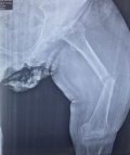
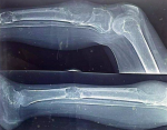
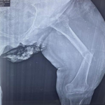
Fourth case: a 70-year-old female patient underwent surgery for secondary hyperparathyroidism, which was diagnosed with disseminated bone pain. Her calcemia measured 73mg/L (85 to 105mg/L), her PTH level was 1761pg/ml (27 times the normal range: 15 to 65 pg/ml), and imaging revealed one left parathyroid nodule, measuring 25mm in its greatest dimension. A parathyroidectomy was performed; the histopathological examination was consistent with a hyperplastic adenoma. We note that all patients presented with radiological evidence of bone disease, brown tumours (Figure 1 and Figure 2), and vitamin D deficiency, and they all received preoperative bisphosphonates, except for the fourth patient.
Clinical findings: clinical examination in the postoperative period showed a positive Chvostek sign and Trousseau sign for all patients. Electrocardiography (ECG) demonstrated QT interval prolongation in one patient, while findings remained within normal limits for the others.
Diagnostic assessment: the postoperative course was marked by the onset of hypocalcemia persisting for more than four days after surgery, hypophosphatemia, and a normal PTH level. A diagnosis of HBS was established.
Therapeutic intervention: all patients had blood calcium below 70mg/l and they were treated with calcium gluconate injection in two phases: attack phase with three ampoules of calcium in 100ml of 5% dextrose solution (D5W) and maintenance phase with five ampoules in 500cc of 5% D5W every twelve hours. This protocol was adapted each time according to blood calcium results. The patients were discharged 45 days on average after parathyroidectomy with the following treatment: oral calcitriol 1µg (two times daily) and calcium carbonate 1,000mg (three times daily).
Follow-up and outcomes: Chvostek's sign and Trousseau's sign were negative for all patients, and they reported no further symptoms. Regarding the biologic tests, blood calcium normalised with oral treatment, and a long-term evaluation is planned.
Consent: all patients provided consent for the surgical intervention.
Patient perspective: all patients expressed satisfaction following the procedure.
The reported prevalence of hungry bone syndrome (HBS) has exhibited significant temporal and geographic variations. While historical data estimated an incidence of approximately 13% following parathyroidectomy for primary hyperparathyroidism, contemporary series often report lower rates, sometimes reaching 4% [1]. Conversely, some Asian cohorts have demonstrated substantially higher frequencies, with prevalence rates peaking at 87%. These discrepancies are likely attributable to differences in healthcare accessibility; earlier diagnosis and prompt surgical intervention for hyperparathyroidism appear to mitigate the risk of developing severe postoperative HBS [2-4].
Multiple risk factors have been identified in retrospective studies, case reports, and case series reviews that correlate with HBS. These factors include elevated levels of PTH, alkaline phosphatase, body mass index, blood urea nitrogen, and the size of resected glands. Moreover, evidence of bone diseases such as brown tumors, fractures, and osteitis fibrosa cystica, along with higher osteoclast counts on bone biopsy, has been correlated with an increased risk of HBS [1]. Indeed, in our case series, all four patients presented elevated PTH levels and significant glandular volume. Additionally, two patients exhibited high alkaline phosphatase levels. Conversely, all four patients had a low Body Mass Index (BMI). The low BMI observed in our patients may be attributed to a higher prevalence of profound vitamin D deficiency and a later diagnosis, leading to more severe bone disease and chronic hypercalcemia-induced anorexia.
The syndrome is reported in 25-90% of patients with radiological evidence of hyperparathyroid bone disease vs only 0-6% of patients without skeletal involvement [5]. Consistently, all cases in our series exhibited radiological evidence of bone disease. Divergent risk factors have been observed in the incidence of HBS. In primary hyperparathyroidism, older age and higher preoperative calcium levels are associated with HBS, while in secondary hyperparathyroidism, younger age and lower preoperative calcium levels have been linked to the condition [2,3]. In our case series, we observed a distinct pattern: patients with primary hyperparathyroidism (PHPT) were younger and presented with higher preoperative calcium levels. Conversely, our cases of secondary hyperparathyroidism (SHPT) involved older patients with lower calcium levels. Regarding the age distribution, the younger presentation of PHPT in our series likely reflects a late-stage diagnosis of more aggressive phenotypes, which become symptomatic earlier in life.
Preoperative treatment with bisphosphonates has been suggested to reduce post-operative hypocalcaemia by several retrospective studies [6]. In a study by Malabu and Founda including 46 patients with severe bone disease, who were treated with intravenous zoledronate preoperatively, the rate of hungry bone syndrome was only 4% [7]. Another retrospective study by França et al. including six patients with radiological features of osteitis fibrosa cystica, who were preoperatively treated with bisphosphonates, reported that none of the patients needed postoperative intravenous calcium supplementation [8]; however, there are to date no prospective studies addressing this issue. The first three patients in our series received preoperative bisphosphonates; nevertheless, they required intravenous calcium administration and subsequently developed hungry bone syndrome (HBS).
For treatment, intravenous calcium replacement, preferably via calcium gluconate due to its lower osmolality and reduced risk of tissue necrosis upon extravasation, is indicated for serum levels below 76mg/L symptomatic hypocalcemia, or QTc interval prolongation. Although calcium chloride provides a higher elemental calcium concentration, calcium gluconate remains the clinical standard as it can be safely administered through peripheral venous access [9,10]. Our patients were also treated with calcium gluconate.
Oral treatment consisted of calcium and magnesium supplementation; the patient should also receive active vitamin D. Calcitriol (0.25-1 mcg/d) can be used, keeping in mind that the effects of vitamin D may take several days to correlate with changes in calcium levels [1]. This same therapeutic regimen was applied to our patient cohort. The long-term outlook for individuals affected by HBS is highly variable, especially regarding the timeline for resolution. Regarding the length of stay, the occurrence of HBS significantly prolongs postoperative hospitalisation compared to uncomplicated parathyroidectomy. In the literature, the mean hospital stay for these patients ranges from 5 to 10 days, though it can extend to several weeks in severe cases, as in our patients. This prolonged duration is primarily dictated by the time required to stabilise serum calcium levels and transition from intensive intravenous supplementation to an effective oral regimen. Clinical evidence indicates that requirements for supplemental calcium and active vitamin D metabolites may extend for as long as twelve months following the surgical procedure.
Although frequently underestimated, hungry bone syndrome (HBS) represents a significant aetiology of hypocalcemia following thyroid or parathyroid surgery. It is imperative that the multidisciplinary clinical team maintains high surveillance for postoperative electrolyte imbalances. Monitoring serum PTH, phosphorus, and magnesium is essential for a precise differential diagnosis. Ultimately, enhanced clinical suspicion and a standardised management approach are vital to optimising patient outcomes and avoiding diagnostic delays.
The authors declare no competing interests.
Chaimaa El Hadri: patient management and drafting of the manuscript. Nada Ait Kassi and Hiba Kouira: data collection. Fatima Toulali and Ghizlane Sabbar: patient management and critical revision of the manuscript. Kaoutar Rifai, Hinde Iraqi and Mohammed El Hassan Gharbi: critical revision of the manuscript. All authors read and approved the final version of the manuscript.
Figure 1: supracondylar fracture
Figure 2: characteristic features of brown tumours
- Cartwright C, Anastasopoulou C. Hungry Bone Syndrome. 2025 Feb 6. In: StatPearls [Internet]. Treasure Island (FL): StatPearls Publishing; 2025 Jan‐. PubMed | Google Scholar
- Witteveen JE, van Thiel S, Romijn JA, Hamdy NA. Hungry bone syndrome: still a challenge in the post-operative management of primary hyperparathyroidism: a systematic review of the literature. Eur J Endocrinol. 2013 Mar;168(3):R45-53. PubMed | Google Scholar
- Jain N, Reilly RF. Hungry bone syndrome. Curr Opin Nephrol Hypertens. 2017 Jul;26(4):250-255. PubMed | Google Scholar
- Brasier AR, Nussbaum SR. Hungry bone syndrome: clinical and biochemical predictors of its occurrence after parathyroid surgery. Am J Med. 1988 Apr;84(4):654-60. PubMed | Google Scholar
- Silva BC, Bilezikian JP. Parathyroid hormone: anabolic and catabolic actions on the skeleton. Curr Opin Pharmacol. 2015 Jun;22:41-50. PubMed | Google Scholar
- Mayilvaganan S, Vijaya Sarathi HA, Shivaprasad C. Preoperative zoledronic acid therapy prevent hungry bone syndrome in patients with primary hyperparathyroidism. Indian J Endocrinol Metab. 2017 Jan-Feb;21(1):76-79. PubMed | Google Scholar
- Malabu UH, Founda MA. Primary hyperparathyroidism in Saudi Arabia: A review of 46 cases. Med J Malaysia. 2007 Dec;62(5):394-7. PubMed | Google Scholar
- França TC, Griz L, Pinho J, Diniz ET, Andrade LD, Lucena CS et al. Bisphosphonates can reduce bone hunger after parathyroidectomy in patients with primary hyperparathyroidism and osteitis fibrosa cystica. Rev Bras Reumatol. 2011 Mar-Apr;51(2):131-7. PubMed | Google Scholar
- Turner J, Gittoes N, Selby P, Society for Endocrinology Clinical Committee. SOCIETY FOR ENDOCRINOLOGY ENDOCRINE EMERGENCY GUIDANCE: Emergency management of acute hypocalcaemia in adult patients. Endocr Connect. 2016 Sep;5(5):G7-G8. PubMed | Google Scholar
- Kelly A, Levine MA. Hypocalcemia in the critically ill patient. J Intensive Care Med. 2013 May-Jun;28(3):166-77. PubMed | Google Scholar
Search
This article authors
On Pubmed
On Google Scholar
Citation [Download]
Navigate this article
Similar articles in
Key words
Tables and figures
Article metrics
Recently from the PAMJ


Authors´ services