
A rare case of long-segment syringomyelia extending from C3 to D9 vertebrae with associated cervical Tarlov cyst in a 52-year-old female: a case report
Madhuri Deotale, Nandini Palamwar, Archana Maheshgauri, Sarika Khadse, Tarun Ajit
Corresponding author: Madhuri Deotale, Department, Smt. Radhikabai Meghe Memorial College of Nursing (FNTCN), Datta Meghe Institute of Higher Education and Research, Wardha, Maharashtra, India 
Received: 06 Jun 2026 - Accepted: 17 Jun 2026 - Published: 26 Aug 2026
Domain: Clinical nursing, Nursing education, Public Health Nursing
Keywords: Syringomyelia, Tarlov cyst, spinal cord, magnetic resonance imaging, neurological disorder, case report
Funding: This work received no specific grant from any funding agency in the public, commercial, or non-profit sectors.
©Madhuri Deotale et al. Pan African Medical Journal (ISSN: 1937-8688). This is an Open Access article distributed under the terms of the Creative Commons Attribution International 4.0 License (https://creativecommons.org/licenses/by/4.0/), which permits unrestricted use, distribution, and reproduction in any medium, provided the original work is properly cited.
Cite this article: Madhuri Deotale et al. A rare case of long-segment syringomyelia extending from C3 to D9 vertebrae with associated cervical Tarlov cyst in a 52-year-old female: a case report. Pan African Medical Journal. 2026;54:143. [doi: 10.11604/pamj.2026.54.143.53800]
Available online at: https://www.panafrican-med-journal.com//content/article/54/143/full
 PDF in process
 Case report 

A rare case of long-segment syringomyelia extending from C3 to D9 vertebrae with associated cervical Tarlov cyst in a 52-year-old female: a case report
A rare case of long-segment syringomyelia extending from C3 to D9 vertebrae with associated cervical Tarlov cyst in a 52-year-old female: a case report
Madhuri Deotale1,&,  Nandini Palamwar1, Archana Maheshgauri1, Sarika Khadse1, Tarun Ajit1
&Corresponding author
Syringomyelia is an uncommon neurological disorder characterized by the formation of a fluid-filled cavity within the spinal cord. Clinical manifestations are often nonspecific, resulting in delayed diagnosis and management. The coexistence of long-segment syringomyelia with a cervical Tarlov cyst is rare and presents diagnostic challenges. A 52-year-old female presented with headache, generalized weakness, and numbness of the left upper limb for one month. She also reported fever with chills for fifteen days. Clinical examination was largely unremarkable except for subjective sensory symptoms involving the left upper limb. Magnetic resonance imaging (MRI) of the cervical spine demonstrated a long-segment syrinx extending from the upper end plate of C3 to the lower end plate of D9 vertebra. A 4 x 4 mm Tarlov cyst was identified within the right neural foramina at the C6-C7 level. Additional findings included early degenerative cervical spine changes, mild central disc bulges, and a posterior annular tear at C5-C6. The patient received symptomatic treatment and underwent neurosurgical evaluation. This case highlights the importance of considering spinal cord pathology in patients presenting with persistent unexplained neurological symptoms. MRI remains the cornerstone of diagnosis, and timely neurosurgical assessment is essential to prevent progression and neurological disability.

Syringomyelia is a chronic neurological disorder characterized by the formation of a fluid-filled cavity within the spinal cord known as a syrinx [1]. Clinical manifestations are often nonspecific and may include pain, sensory disturbances, weakness, and autonomic dysfunction, frequently leading to delayed diagnosis [2]. The condition may occur secondary to Chiari malformation, spinal cord tumors, trauma, or may be idiopathic in origin [3].
Magnetic resonance imaging (MRI) is considered the gold standard for diagnosis because it allows detailed visualization of the spinal cord and associated abnormalities [4]. Tarlov cysts are cerebrospinal fluid-filled dilatations of nerve root sheaths that are usually asymptomatic but may occasionally produce neurological symptoms due to nerve root compression [5].
We report a rare case of extensive syringomyelia extending from C3 to D9 vertebral levels associated with a cervical Tarlov cyst at the C6-C7 level in a 52-year-old female.

Patient information: a 52-year-old female presented to the medicine outpatient department with complaints of headache, generalized weakness, and numbness involving the left upper limb for one month. She also experienced fever with chills for fifteen days before admission.
The symptoms developed gradually and were persistent. The patient reported worsening discomfort on prolonged standing and partial relief with rest. There was no history of trauma, loss of consciousness, seizures, syncope, bowel or bladder dysfunction, chest pain, palpitations, or respiratory symptoms.
Past medical history was negative for hypertension, diabetes mellitus, tuberculosis, thyroid disorders, bronchial asthma, previous spinal surgery, or known neurological disease. Family history was non-contributory.
Clinical findings: on examination, the patient was conscious, alert, cooperative, and oriented to time, place, and person. Vital signs were stable: pulse rate: 80 beats/minute; blood pressure: 120/80 mmHg; respiratory rate: 14 breaths/minute; temperature: afebrile. General physical examination did not reveal pallor, cyanosis, clubbing, edema, or lymphadenopathy. Neurological assessment revealed subjective numbness involving the left upper limb. No gross motor deficit was documented. Cranial nerve examination was normal. Cardiovascular, respiratory, abdominal, and musculoskeletal examinations were unremarkable.
Timeline: approximately one month before admission, the patient developed headache, generalized weakness, and numbness of the left upper limb, which gradually progressed and persisted. Fifteen days before admission, she experienced fever associated with chills. On admission, a comprehensive clinical examination and routine laboratory investigations were performed. During hospitalization, magnetic resonance imaging (MRI) of the brain and cervical spine was conducted, revealing a long-segment syrinx extending from the upper end plate of C3 to the lower end plate of D9 vertebral levels along with a cervical Tarlov cyst at the C6-C7 level. Following confirmation of the diagnosis, a neurosurgical consultation was obtained, and further imaging along with surgical evaluation was advised. The patient remained clinically stable during hospitalization, showed symptomatic improvement with conservative management, and was discharged with recommendations for neurological follow-up and continued neurosurgical assessment.
Diagnostic assessment: routine laboratory investigations, including complete blood count, coagulation profile, erythrocyte sedimentation rate, C-reactive protein, and vitamin B12 levels, were within normal limits. Chest radiography, electrocardiography, and Mantoux testing were unremarkable. MRI of the brain revealed no abnormalities.
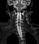
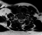
Magnetic resonance imaging of the cervical spine demonstrated a T2 hyperintense syrinx extending from the upper end plate of C3 to the lower end plate of D9 vertebral levels, consistent with long-segment syringomyelia (Figure 1). A T2 hyperintense cystic lesion measuring approximately 4 x 4 mm was identified within the right neural foramina at the C6-C7 level, suggestive of a Tarlov cyst (Figure 2). Additional findings included mild central disc bulges at C3-C4, C4-C5, and C5-C6 levels, a posterior annular tear at C5-C6, and early degenerative cervical spine changes. Whole-spine screening demonstrated posterior disc bulge and annular tear at L5-S1.
Diagnostic challenges: the patient's symptoms were initially nonspecific and suggestive of a musculoskeletal or inflammatory disorder. The absence of significant neurological deficits contributed to diagnostic uncertainty. Definitive diagnosis was achieved only after MRI evaluation.
Diagnosis: long-segment syringomyelia extending from C3 to D9 vertebral levels associated with a cervical Tarlov cyst at the C6-C7 level.
Therapeutic intervention: the patient was admitted for further evaluation and symptomatic management. During hospitalization, she received intravenous ceftriaxone and oral azithromycin for the management of fever and associated symptoms. Supportive medications included levocetirizine for symptomatic relief and hydroxychloroquine as prescribed by the treating physician. Neuropathic symptoms, particularly numbness of the left upper limb, were managed with gabapentin. The patient was closely monitored for neurological deterioration, progression of sensory symptoms, and changes in vital parameters.
Following radiological confirmation of long-segment syringomyelia extending from C3 to D9 vertebral levels with an associated Tarlov cyst at the C6-C7 level, a neurosurgical consultation was obtained. Further evaluation with MRI of the lumbosacral spine was recommended to exclude associated tethered cord syndrome. The neurosurgical team advised consideration of surgical intervention, including foramen magnum decompression with cord detethering if clinically indicated. The patient remained hemodynamically stable throughout hospitalization and demonstrated symptomatic improvement with conservative management.
Follow-up and outcomes: the patient remained hemodynamically stable throughout hospitalization. Symptoms improved with conservative treatment. She was discharged in stable condition with advice regarding medications, neurological monitoring, and neurosurgical follow-up for further evaluation and definitive management planning.
Patient perspective: the patient expressed relief after receiving a definitive diagnosis for her symptoms and agreed to continue further evaluation and follow-up. Written informed consent was obtained for publication of this case report and accompanying images.
Patient consent: written informed consent was obtained from the patient for publication of this case report and accompanying images.
The present case demonstrates a rare association between long-segment syringomyelia and a cervical Tarlov cyst. Syringomyelia often presents with subtle and nonspecific symptoms, which may delay diagnosis and treatment [6]. In the present patient, headache, generalized weakness, and upper limb numbness initially suggested a non-neurological condition, highlighting the importance of maintaining a broad differential diagnosis.
Magnetic resonance imaging remains the investigation of choice because it accurately identifies the extent of the syrinx and associated abnormalities [4]. The extensive involvement from C3 to D9 vertebral levels observed in this patient is uncommon and may increase the risk of neurological impairment due to widespread spinal cord involvement [7].
Tarlov cysts are most frequently encountered in the sacral region and are often discovered incidentally. However, symptomatic cysts can contribute to neurological manifestations and complicate clinical management [8]. The coexistence of syringomyelia and a cervical Tarlov cyst has rarely been reported in the literature.
Early neurosurgical consultation is essential because progressive enlargement of a syrinx may lead to irreversible neurological deficits. Surgical treatment is often directed toward correction of cerebrospinal fluid flow abnormalities and prevention of disease progression [9].
The diagnosis was confirmed by MRI, which clearly demonstrated the extensive syrinx extending from C3 to D9 vertebral levels (Figure 1) and the associated cervical Tarlov cyst at the C6-C7 level (Figure 2).
Long-segment syringomyelia extending from C3 to D9 vertebral levels associated with a cervical Tarlov cyst is an uncommon clinical entity. The nonspecific nature of presenting symptoms may delay diagnosis. MRI plays a pivotal role in identifying the lesion and associated abnormalities. Early diagnosis, multidisciplinary management, and timely neurosurgical evaluation are essential for preventing neurological deterioration and improving patient outcomes.
The authors declare no competing interests.
The authors declare no competing interests.
Figure 1: coronal T2-weighted magnetic resonance image of the cervical and upper thoracic spine showing a long-segment syrinx extending from the upper end plate of C3 to the lower end plate of D9 vertebral levels
Figure 2: axial T2-weighted magnetic resonance image at the C6-C7 level demonstrating a 4 x 4 mm Tarlov cyst within the right neural foramina
- NHI. Syringomyelia. 2026. Accessed 23rd April, 2026.
- Mayo clinic. Diseases & Conditions: Syringomyelia: Symptoms & causes. Mayo Clinic. 2026. Accessed 23rd April, 2026.
- Toader C, Ples H, Covache-Busuioc RA, Costin HP, Bratu BG, Dumitrascu DI et al. Decoding Chiari Malformation and Syringomyelia: From Epidemiology and Genetics to Advanced Diagnosis and Management Strategies. Brain Sci. 2023 Nov 30;13(12):1658. PubMed | Google Scholar
- Mayo clinic. Diseases & Conditions: Syringomyelia: Diagnosis & treatment. Mayo Clinic. 2026. Accessed 23rd April, 2026.
- Lim Y, Selbi W. Tarlov Cyst. In: StatPearls [Internet]. Treasure Island (FL): StatPearls Publishing; 2026 Jan. 2023. PubMed | Google Scholar
- Shenoy VS, Munakomi S, Sampath R. Syringomyelia. In: StatPearls [Internet]. Treasure Island (FL): StatPearls Publishing; 2026 Jan. 2024. PubMed | Google Scholar
- Fadila M, Sarrabia G, Shapira S, Yaacobi E, Baruch Y, Engel I et al. Orthopedic Manifestations of Syringomyelia: A Comprehensive Review. J Clin Med. 2025 May 1;14(9):3145. PubMed | Google Scholar
- Murphy K, Nasralla M, Pron G, Almohaimede K, Schievink W. Management of Tarlov cysts: an uncommon but potentially serious spinal column disease-review of the literature and experience with over 1000 referrals. Neuroradiology. 2024 Jan;66(1):1-30. PubMed | Google Scholar
- Bhagavathula Venkata SS, Arimappamagan A, Lafazanos S, Pruthi N. Syringomyelia secondary to cervical spondylosis: Case report and review of literature. J Neurosci Rural Pract. 2014 Nov;5(Suppl 1):S78-82. PubMed | Google Scholar
Search
This article authors
On Pubmed
On Google Scholar
Citation [Download]
Navigate this article
Similar articles in
Key words
Tables and figures
Article metrics
Recently from the PAMJ


Authors´ services
