
Recurrent sinonasal inverted papilloma and the importance of complete excision: a case report
Amoghvarsh Mallikarjun Jamadar, Prasad Deshmukh, Sagar Gaurkar, Himanshi Kesharwani, Srushti Patel
Corresponding author: Amoghvarsh Mallikarjun Jamadar, Datta Meghe Institute of Higher Education and Research, Sawangi, Wardha, Maharashtra, India 
Received: 18 Mar 2026 - Accepted: 04 May 2026 - Published: 26 Aug 2026
Domain: Otolaryngology (ENT)
Keywords: Endoscopic surgery, maxillary sinus, nasal cavity, recurrence, schneiderian papilloma, case report
Funding: This work received no specific grant from any funding agency in the public, commercial, or non-profit sectors.
©Amoghvarsh Mallikarjun Jamadar et al. Pan African Medical Journal (ISSN: 1937-8688). This is an Open Access article distributed under the terms of the Creative Commons Attribution International 4.0 License (https://creativecommons.org/licenses/by/4.0/), which permits unrestricted use, distribution, and reproduction in any medium, provided the original work is properly cited.
Cite this article: Amoghvarsh Mallikarjun Jamadar et al. Recurrent sinonasal inverted papilloma and the importance of complete excision: a case report. Pan African Medical Journal. 2026;54:141. [doi: 10.11604/pamj.2026.54.141.52261]
Available online at: https://www.panafrican-med-journal.com//content/article/54/141/full
 PDF in process
 Case report 

Recurrent sinonasal inverted papilloma and the importance of complete excision: a case report
Recurrent sinonasal inverted papilloma and the importance of complete excision: a case report
 Amoghvarsh Mallikarjun Jamadar1,&, Prasad Deshmukh1, Sagar Gaurkar1, Himanshi Kesharwani1, Srushti Patel1
&Corresponding author
Sinonasal inverted papilloma (SIP), a benign epithelial tumor, typically grows on the nasal cavity mucosa and on the paranasal sinuses. They grow rapidly, recur frequently, and can progress to a malignant form. This case reports a 74-year-old male with right nasal obstruction, intermittent epistaxis, and nasal discharge. He had previously undergone a nasal papilloma removal. Endoscopic and radiographic evaluation revealed the recurrence of the disease in the right nasal cavity involving the maxillary and ethmoidal sinuses. Histopathologic analysis revealed SIP. He underwent an endoscopic procedure for tumor removal and separation of scarring using a coblator and microdebriders through a lateral rhinotomy approach. The postoperative recovery was uncomplicated, and an endoscopy done one month later showed complete healing with no residual. This case emphasizes the need for total surgical removal of the tumor and continued endoscopic examinations to monitor for recurrence and transformation to malignancy.

Sinonasal inverted papilloma (SIP) is a rare benign epithelial tumor of the nasal cavity and paranasal sinuses. It typically grows locally but tends to recur after initial surgical excision. Symptoms include unilateral nasal obstruction, epistaxis, and nasal discharge. Sinonasal inverted papilloma (SIP) can be misdiagnosed for common upper respiratory problems such as chronic rhinosinusitis or nasal polyps [1]. Considered benign based on histology, an association with malignancy has been reported. Due to the aggressive nature and potential for malignant transformation, careful surgical planning and total excision of the tumor are critical to minimizing recurrence. Long-term follow-up care with routine endoscopic evaluations is necessary to evaluate for recurrence and the development of malignancy [2]. We report the case of a 74-year-old male who had multiple episodes of nasal obstruction and epistaxis for 6 years. Imaging confirmed the diagnosis of SIP, surgical removal was performed, short-term follow-up showed no residual disease, and given the high recurrence rate, long-term follow-up was recommended.

Patient information: a 74-year-old male, previously treated for right-sided nasal squamous papilloma 6 years ago, with no regular follow-up, otherwise with no significant systemic abnormalities on evaluation.
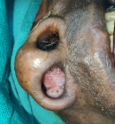
Clinical findings: the patient presented with progressive right-sided nasal obstruction, mucoid nasal discharge, and intermittent epistaxis. The obstruction began insidiously 2 years ago, initially intermittent and later becoming continuous over the past 4 months (Figure 1). Epistaxis increased from once or twice monthly to 2-4 times weekly and was self-limiting. He also reported hyposmia, mouth breathing during sleep, snoring, and sleep disturbances. There were no associated symptoms such as facial pain, headache, visual disturbances, hearing loss, dysphagia, voice changes, fever, or weight loss. Anterior rhinoscopy revealed a fleshy pinkish polypoid mass filling the right nasal cavity with an irregular surface and bleeding on palpation, along with deviation of the nasal septum to the left. The left nasal cavity was normal.
Timeline of current episode: six years prior: excision of right nasal mass diagnosed as squamous papilloma. Two years prior: onset of intermittent nasal obstruction. Four months prior: progression to continuous obstruction with increased frequency of epistaxis. At presentation: diagnostic workup and imaging performed. Post-surgery: nasal packing removed on day 2. One-month follow-up: no residual symptoms or disease.
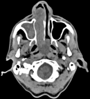
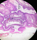
Diagnostic assessment: diagnostic nasal endoscopy showed a firm lobulated mass in the right nasal cavity extending beyond the choana, obscuring the middle turbinate and osteomeatal complex. Contrast-enhanced computed tomography revealed a large heterogeneous polyp (7.3 x 3.5 x 4.1 cm) occupying the right nasal cavity and extending into the right maxillary and ethmoid sinuses, without intracranial or orbital extension, consistent with SIP (Figure 2). Laboratory investigations, including blood count, coagulation profile, liver and kidney function tests, thyroid function, and viral serology, were normal. Histopathology showed no dysplasia or malignancy, and immunohistochemistry confirmed SIP without malignant transformation (Figure 3).
Diagnosis: SIP involving the right nasal cavity, maxillary sinus, and ethmoid sinus.
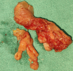
Therapeutic interventions: right endoscopic combined endonasal coblation microdebrider-assisted maxillary sinus surgery with lateral rhinotomy. Complete excision of the tumor was performed using a microdebrider, with stripping of maxillary sinus mucosa and osteotomies for full access (Figure 4). The middle turbinate was preserved and repositioned. Image guidance was used intraoperatively, and adequate hemostasis was achieved with nasal packing.
Follow-up and outcome of interventions: postoperative recovery was uneventful. At 1-month follow-up, the patient was asymptomatic with no nasal obstruction, epistaxis, or discharge. Endoscopic evaluation showed a healthy nasal cavity with no evidence of residual or recurrent disease, and improvement in sense of smell was noted. Long-term follow-up with periodic endoscopic evaluation for up to 5 years was advised due to the risk of recurrence.
Patient perspective: for the past two years, my continuous right-sided nasal blockage, frequent nosebleeds, and loss of smell made daily life and sleeping incredibly frustrating. Having had a similar growth removed six years ago without maintaining my follow-up appointments, I was anxious when the symptoms returned and rapidly worsened. Fortunately, despite needing a more extensive surgery this time to remove the recurrent tumor, my recovery was surprisingly smooth. Just one month post-surgery, my breathing is completely clear, my sense of smell has returned, and the bleeding has stopped entirely. This experience has been a true wake-up call, and I am now fully committed to attending all my routine endoscopic check-ups over the next five years to ensure it does not come back.
Informed consent: informed consent was obtained.
Sinonasal inverted papilloma is a fairly uncommon, benign tumor that develops in the sinuses and represents between 0.4-4.7% of all tumors of this type. The estimated rate of occurrence ranges from 0.2 to 1.5 cases per 100,000 people per year [3]. Sinonasal inverted papilloma (SIP) is more common in men, and typically occurs between the ages of 50 and 70 years. The most common symptoms include unilateral nasal obstruction, nasal drainage, and occasional epistaxis [1]. Sinonasal inverted papilloma (SIP) most frequently starts in either the maxillary or ethmoid sinuses, but it can be found in adjacent structures. While SIP does not spread throughout the body like malignant tumors, it is an aggressive tumor that has a recurrence rate of 5-20%, and the chance of malignant transformation is about 6-10% [3]. Treatment for SIP generally includes surgical removal, and whether or not the patient has a good outcome from the surgery is directly dependent on whether or not the entire tumor has been removed completely, and if there is subsequent follow-up care, since recurrences can occur years after the initial surgery [4].
Treatment plans for patients with SIPs should be tailored to each patient. Olsson et al. documented a patient with an SIP of the posterior ethmoid who underwent successful image-assisted endoscopic surgery. Olsson et al. emphasized the need to accurately identify and dissect the base of inverted papillomas to maximize surgical success and minimize recurrence [5]. Our case demonstrated the importance of identifying exactly where the SIP was attached to adjacent structures, despite the very diffuse extent of disease.
Basavanniah et al. concluded that imaging and clinical assessment are both essential for establishing a diagnosis of an "antrochoanal-like" lesion. However, histology of the biopsy specimen is more accurate than either imaging or clinical evaluation in establishing a definitive diagnosis [6]. Dudda et al. performed endoscopic medial maxillectomy in patients with multiple sinuses involved by the disease. They reported that appropriately employed endoscopic surgical techniques can be successfully used for extensive disease and result in less morbidity than traditional surgical techniques [7]. Khandekar et al. suggested that an incompletely excised tumor at the time of initial evaluation, due to a lack of histologic evidence, may lead to tumor recurrence, requiring an open procedure to completely excise the tumor [8].
Bilateral SIPs can be challenging because they are not typical findings, as noted by Al-Momen et al. who performed navigational endoscopic medial maxillectomy in patients with bilateral inverted papilloma. Their technique resulted in good long-term outcomes. Because there is a difference in surgical outcome quality between endoscopically and open surgical procedures, it stands to reason that a poor-quality surgical outcome would increase the risk of tumor recurrence or persistence [2].
Ongoing histopathologic assessments, along with diligent follow-up, are required for individuals with these high-risk characteristics. Based on their findings, Eggers et al. showed that there is an association between increased mitotic activity, dyskaryosis, male gender, and advancing age and the risk of malignancy. Their recommendation was to obtain en bloc histological specimens and to sample tissue in a non-fragmented, complete manner whenever attainable [9].
Endoscopic technique and surgical intervention in the treatment of sinonasal neoplastic disease are the most frequent and common options for the management of these neoplasms due to their lower morbidity and increased surgical visualization. There are situations in which an open or combined surgical approach may provide the best solution for patients with recurrent, extensive, or complex sinonasal disease. The extent of the disease should determine the surgical strategy rather than a uniform approach. Long-term endoscopic follow-up of all patients is recommended to assess the recurrence of neoplastic lesions and the risk of malignancy [5,7].
In this elderly patient with SIP, the disease presented as an extensive, recurrent unilateral lesion in the nasal cavity and paranasal sinuses and responded favorably to a combined open and endoscopic surgical approach. Patients with prior sinonasal surgery and recurrence of symptoms require thorough preoperative evaluation, a high degree of suspicion for recurrence or malignant transformation, complete surgical removal of the tumor attachment site using a modified combined surgical technique and powered instrumentation to prevent recurrence and ensure positive long-term outcomes through continued structured endoscopic follow-up.
The authors declare no competing interests.
Conceptualization: Amoghvarsh Mallikarjun Jamadar and Prasad Deshmukh; data curation: Amoghvarsh Mallikarjun Jamadar and Sagar Gaurkar; format analysis: Amoghvarsh Mallikarjun Jamadar, Prasad Deshmukh, and Himanshi Kesharwani; investigation: Amoghvarsh Mallikarjun Jamadar and Srushti Patel; methodology: Sagar Gaurkar and Himanshi Kesharwani; writing final draft: Amoghvarsh Mallikarjun Jamadar; writing-reviewing and editing: all authors. All the authors read and approved the final version of this manuscript.
Figure 1: clinical image showing a lobulated pinkish-red mass occluding the right nasal vestibule and anterior nasal cavity
Figure 2: axial contrast-enhanced computed tomography showing expansile, enhancing soft tissue mass in the right nasal cavity extending into right maxillary sinus laterally, superiorly into right ethmoidal air cells and posteriorly into nasopharynx with marked contralateral septal deviation
Figure 3: H&E (4x): endophytic invagination of thickened Schneiderian epithelium into the stroma (black arrow) with preserved polarity and absence of dysplasia (red arrow)
Figure 4: gross surgical specimen showing excised lobulated soft tissue mass in multiple fragments
- Upadhya IB, Rao K. Sinonasal Inverted Papilloma: A Narrative Review. Indian J Otolaryngol Head Neck Surg. 2022 Oct;74(Suppl 2):1017-1022. PubMed | Google Scholar
- Al Momen A, Alenzi HL, Al Eid M. Bilateral simultaneous sino-nasal inverted papilloma; A report of two cases and literature review. Int J Surg Case Rep. 2020;67:71-75. PubMed | Google Scholar
- Li X, Wang D, Yin D. Clinical characteristics and recurrence predictors of sinonasal inverted papilloma: a retrospective cohort study. Front Oncol. 2025 Apr 7;15:1537905. PubMed | Google Scholar
- Gutiérrez-Posso JD, Zabala-López-de-Maturana A, Arango-Molina AM, Sagazola-Odriozola J, Santaolalla-Sánchez FJ. Sinonasal inverted papilloma surgery over two decades: Outcomes and evolution of practice. Rev ORL. 2025;16(4):e33565. Google Scholar
- Olsson SE, Peña R. Endoscopic Removal of Sinonasal Inverted Papilloma Originating From the Posterior Ethmoid Cavity. Case Rep Otolaryngol. 2025 Feb 20;2025:8389174. PubMed | Google Scholar
- Basavannaiah S. Tricky nasal mass from meek polyp to intricate papilloma: A diagnostic dilemma. IP J Otorhinolaryngol Allied Sci. 2021;4(3):115-118.
- Dudda R, Jayanna SH, M HP, Balaji NK. Endoscopic resection of inverted papilloma of nasal cavity: A case report. Int J Otorhinolaryngol Head Neck Surg. 2018;4(3):846-849.
- Khandekar S, Dive A, Mishra R, Upadhyaya N. Sinonasal inverted papilloma: A case report and mini review of histopathological features. J Oral Maxillofac Pathol. 2015 Sep-Dec;19(3):405. PubMed | Google Scholar
- Eggers G, Eggers H, Sander N, Kössling F, Chilla R. Histological features and malignant transformation of inverted papilloma. Eur Arch Otorhinolaryngol. 2005 Apr;262(4):263-8. PubMed | Google Scholar
Search
This article authors
On Pubmed
On Google Scholar
Citation [Download]
Navigate this article
Similar articles in
Key words
Tables and figures
Article metrics
Recently from the PAMJ


Authors´ services
