
Pleural biphasic synovial sarcoma: a case report
Rahul Shil, Shreyasee Das, Saikat Das
Corresponding author: Rahul Shil, Department of Neuroscience, People's Education Society University, Bengaluru, India 
Received: 20 Apr 2025 - Accepted: 19 Jun 2026 - Published: 05 Aug 2026
Domain: Radiology, Neuro-oncology
Keywords: Sarcoma, chest wall tumors, malignant pleural mesothelioma, pleural diseases, hemorrhagic pleural effusion, case report
Funding: This work received no specific grant from any funding agency in the public, commercial, or non-profit sectors.
©Rahul Shil et al. Pan African Medical Journal (ISSN: 1937-8688). This is an Open Access article distributed under the terms of the Creative Commons Attribution International 4.0 License (https://creativecommons.org/licenses/by/4.0/), which permits unrestricted use, distribution, and reproduction in any medium, provided the original work is properly cited.
Cite this article: Rahul Shil et al. Pleural biphasic synovial sarcoma: a case report. Pan African Medical Journal. 2026;54:111. [doi: 10.11604/pamj.2026.54.111.47659]
Available online at: https://www.panafrican-med-journal.com//content/article/54/111/full
 PDF in process
 Case report 

Pleural biphasic synovial sarcoma: a case report
Pleural biphasic synovial sarcoma: a case report
 Rahul Shil1,&, Shreyasee Das2, Saikat Das3
&Corresponding author
Synovial sarcoma is a rare and aggressive soft tissue malignancy that typically arises near joint cavities but can occasionally occur in unusual locations such as the pleura. Pleural synovial sarcoma is extremely uncommon and often presents with nonspecific symptoms such as chest pain, dyspnea, and weight loss, which can mimic more common pleural conditions like mesothelioma or tuberculosis. Due to its rarity and overlapping clinical features, diagnosis often requires advanced imaging, histopathology, and immunohistochemistry for confirmation. This case study describes a young adult patient diagnosed with pleural synovial sarcoma and highlights the diagnostic challenges, treatment interventions, and the importance of a multidisciplinary approach in managing this rare malignancy.

Pleural synovial sarcoma is a very rare disease. It is a pernicious soft tissue tumour that most probably occurs in the pleural cavity. This most commonly affects the proximal areas of the large joints [1]. They do not make an appearance from the synovial tissues, as the name quite suggests, but they come to light from the multipoint mesenchymal cells. It is commonly a high-grade sarcoma that occurs para-articular to the location of the joints. The most common sites of origin are the thigh, knee, ankle, foot, and upper extremities. This malignancy mainly occurs in adolescents in the age group between 15 and 40 years of age [2,3]. It accounts for only 0.1%-0.5% among all primary lung malignancies. It is a very highly aggressive tumour and also has a very high chance of recurrence. The prognosis rate is very poor for this condition [4]. To know more about the tumour, imaging plays a very vital role [5]. Patients in this condition present with symptoms of cough, chest pain, and dyspnea. The radiography of the chest results in a sharply marginated mass with a static haziness. Computed tomography images show a confined, diverse, magnified abrasion without the involvement of the bone and without the coagulation, excluding the case of the chest wall primary tumour. Magnetic resonance imaging provides the loftier substantiation of nodular soft tissue and many fluid-filled internal components of PPSS. The most probable treatment of PSSS consists of surgical resection followed by chemotherapy, radiation therapy, or both [3,5]. In this study, we report a case of a 55-year-old female who presented with a chief complaint of fever and weakness, including watery eyes.

Patient information: a 55-year-old female was admitted to the department of oncology on 2nd November 2024 with the complaint of fever, weakness, and watery eyes, and also with a previous medical history of status post (S/P) right anterolateral thoracotomy-WLE, followed by recurrence. Later on, she was provisionally diagnosed with a pleura biphasic synovial sarcoma.
Clinical findings: the patient presented with chest pain, dyspnea, and significant weight loss (from 55 kg to 46 kg), indicating a progressive disease course leading to imaging studies that confirmed the presence of pleural synovial sarcoma. A biopsy revealed undifferentiated spindle cell sarcoma, confirming the diagnosis.
Timeline of current episode: symptoms of pleural synovial sarcoma developed over three months, and the patient underwent chemotherapy shortly after the diagnosis.
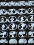
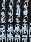
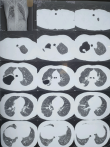
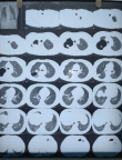
Diagnostic assessment: the investigations for the patient with suspected pleural malignancy were carried out systematically, beginning with imaging and progressing to histopathological confirmation (Figure 1). The patient presented with clinical features such as chest pain, dyspnea, and weight loss, prompting an initial chest X-ray, which revealed a large opacity in the left hemithorax with associated volume loss. This finding raised suspicion for a pleural-based lesion, leading to a contrast-enhanced computed tomography (CECT) scan of the thorax (Figure 2). The CT scan showed a recurrent, heterogeneously enhancing pleural mass on the left side with associated pleural effusion and mediastinal shift, strongly suggestive of a malignant pleural tumour. Further laboratory investigations included routine blood tests, which indicated anaemia and possible signs of inflammation. A pleural fluid analysis was conducted and revealed a hemorrhagic, exudative effusion; however, cytological examination of the fluid was inconclusive in establishing a definitive diagnosis, as is often the case with synovial sarcoma. To confirm the diagnosis, a pleural biopsy was performed, and histopathological examination revealed features consistent with a monophasic spindle cell neoplasm (Figure 3). To differentiate this from other pleural tumours, immunohistochemistry (IHC) was performed, which showed positivity for TLE1, BCL-2, EMA, and focal cytokeratin, while being negative for CD34, S-100, WT-1, and calretinin. This immune profile is characteristic of synovial sarcoma and helped to rule out other differential diagnoses such as mesothelioma or solitary fibrous tumour. Although molecular testing (such as FISH or RT-PCR for the SS18-SSX gene fusion) was not done in this case due to resource constraints, the diagnosis of pleural synovial sarcoma was supported by the clinical findings, imaging, histopathology, and a specific IHC pattern (Figure 4). This structured and multimodal approach ensured accurate diagnosis and guided the subsequent management plan for the patient.
Diagnosis: pleura biphasic synovial sarcoma.
Therapeutic intervention: the treatment course of the patient diagnosed with pleural synovial sarcoma (PSS) followed a multidisciplinary approach involving surgical management, chemotherapy, supportive care, and close follow-up, tailored to the patient's condition and the recurrent nature of the disease. Initially, the patient presented with respiratory symptoms including chest pain, dyspnea, and weight loss. A contrast-enhanced CT scan revealed a recurrent pleural mass with pleural effusion, and histopathological evaluation confirmed a monophasic synovial sarcoma, with immunohistochemistry (IHC) markers such as TLE1, BCL-2, and EMA positive, and WT-1 and calretinin negative, ruling out mesothelioma. Given the recurrence and localised disease, the first step in treatment was surgical resection of the pleural tumour. The goal of surgery was complete tumour excision, but due to pleural adhesions and previous interventions, achieving negative surgical margins was challenging, and microscopic residual disease was suspected postoperatively. Following surgery, the patient was initiated on adjuvant chemotherapy. The regimen included ifosfamide and doxorubicin, which is the standard for soft tissue sarcomas, especially synovial sarcoma, due to its moderate chemosensitivity. The patient tolerated multiple cycles of chemotherapy under close supervision, with routine blood tests to monitor for myelosuppression, renal function, and hepatotoxicity. Throughout the chemotherapy course, supportive care was provided, including antiemetics, analgesics, and nutritional support, to manage chemotherapy side effects and cancer-associated cachexia. In light of the incomplete resection and high risk of local recurrence, radiotherapy was considered for better local control. A course of external beam radiotherapy (EBRT) was planned to the thoracic region to target any residual microscopic disease in the pleura and chest wall. However, due to the patient's declining general condition and side effects from chemotherapy, radiotherapy initiation was postponed, and further evaluation was advised based on treatment response. Throughout the treatment course, the patient was closely monitored for treatment response and symptom management. Repeat imaging was planned after chemotherapy cycles to assess tumour regression. The care plan also included palliative and psychological support, focusing on quality of life and symptom relief, given the chronic and recurrent nature of the disease. In summary, the patient's treatment course included surgical resection, followed by systemic chemotherapy with ifosfamide and doxorubicin, supportive symptom management, and a plan for adjuvant radiotherapy, with regular clinical and radiological follow-up to guide ongoing care and monitor disease progression.
Follow-up and outcome of intervention: after six months, the patient was stable and responded to the treatment a little. However, palliative care and psychological care are still ongoing, but the patient retained walking autonomy.
Patient perspective: the patient expressed shock and grief over her initial symptoms, the diagnosis and how the disease has significantly affected the quality of life.
Informed consent: informed consent was obtained from the patient and family members for the publication of this case report.
Pleural synovial sarcoma is an extremely rare malignancy, constituting less than 1% of all primary lung and pleural tumours [6]. It typically presents with nonspecific symptoms such as chest pain, dyspnea, and pleural effusion, which often lead to a delayed diagnosis [7]. The current case demonstrates the aggressive nature of the disease, with tumour recurrence post-surgical excision, requiring systemic chemotherapy.
Diagnosis and challenges in pleural synovial sarcoma: histopathological evaluation remains the gold standard for diagnosis. Immunohistochemistry plays a crucial role, as pleural synovial sarcomas often express epithelial and mesenchymal markers, including TLE1, BCL2, and EMA [8]. In this case, histopathological analysis confirmed an undifferentiated spindle cell sarcoma, aligning with typical synovial sarcoma features. One of the major challenges in diagnosing pleural synovial sarcoma is distinguishing it from other pleural malignancies, such as malignant mesothelioma and metastatic sarcomas [9]. Cytogenetic testing for SS18 gene rearrangements can confirm the diagnosis, but it is not always available in resource-limited settings [8,9].
Chemotherapy and treatment response: chemotherapy remains the primary treatment modality for inoperable and recurrent synovial sarcomas. The combination of ifosfamide and doxorubicin is considered the first-line therapy due to its efficacy in controlling disease progression [7]. In this case, the patient received four cycles of ifosfamide and doxorubicin, with a partial response noted after Cycle 4. However, the presence of recurrence despite chemotherapy is concerning, as pleural synovial sarcomas have a high rate of local recurrence and metastasis [5]. Studies suggest that dose intensification with ifosfamide or the addition of radiotherapy may improve outcomes in high-risk cases [6,7]. Despite chemotherapy, the patient experienced significant weight loss (55 kg to 46 kg), which is indicative of chemotherapy-induced toxicity and possible disease progression. Weight loss in sarcoma patients is often associated with poor prognosis, necessitating nutritional support and dose adjustments [8].
Alternative and emerging treatment strategies: given the high recurrence rate and poor long-term survival with chemotherapy alone, emerging treatment strategies include:
Targeted therapy: Tyrosine kinase inhibitors (TKIs) such as pazopanib have shown promising results in advanced soft tissue sarcomas.
Immunotherapy: checkpoint inhibitors (PD-1/PD-L1 inhibitors) have been investigated, though synovial sarcomas exhibit low immunogenicity, limiting their efficacy [8,3]. However, combination strategies with immune checkpoint inhibitors and targeted therapy are currently being explored.
Radiotherapy: postoperative radiotherapy is recommended in high-risk synovial sarcoma cases to prevent recurrence [5,6]. In this case, radiotherapy may be considered as an adjuvant therapy to improve local control.
Prognostic factors and long-term outlook: survival in pleural synovial sarcoma is highly variable, with 5-year survival rates ranging from 20% to 60%, depending on factors such as tumour size, completeness of resection, and response to chemotherapy [1,2]. The poor response to chemotherapy in recurrent cases suggests that alternative systemic therapies should be considered early in disease management. This case highlights the aggressive nature of pleural synovial sarcoma, the limitations of chemotherapy, and the need for a multimodal approach incorporating radiotherapy, targeted therapy, and immunotherapy for improved patient outcomes.
Pleural synovial sarcoma is an exceptionally rare and aggressive malignancy that poses significant diagnostic and therapeutic challenges. In this case, the patient presented with nonspecific respiratory symptoms and was eventually diagnosed through a combination of imaging, histopathology, and immunohistochemistry, which confirmed a monophasic synovial sarcoma arising from the pleura. Despite the rarity of this tumour, timely recognition and a multidisciplinary treatment approach were critical in managing the disease. Surgical excision was undertaken as the initial step; however, due to the tumour's recurrent nature and anatomical complexity, complete resection was difficult. This necessitated the use of adjuvant chemotherapy with ifosfamide and doxorubicin, and radiotherapy was considered for further local control. The patient's course of treatment emphasised not only oncologic management but also comprehensive supportive care, focusing on symptom relief and quality of life. This case underscores the importance of early diagnosis, the role of immunohistochemistry in differentiating pleural tumours, and the need for a coordinated treatment strategy to improve outcomes in such rare and challenging malignancies.
The authors declare no competing interests.
Data collection: Saikat Das. Manuscript preparation: Rahul Shil, Shreyasee Das. Patient management and manuscript revision: Rahul Shil, Shreyasee Das and Saikat Das. All authors read and approved the final version of the manuscript.
Figure 1: an axial CT scan of the thorax highlights the different anatomical levels of the chest, including the lungs, heart, and surrounding structures
Figure 2: sagittal and coronal sections providing detailed cross-sectional analysis of the thoracic cavity
Figure 3: axial CT scans of the lungs show lung parenchyma with abnormalities suggestive of pleural involvement
Figure 4: pleural synovial sarcoma indicators visible
- Colwell AS, D’Cunha J, Vargas SO, Parker B, Di Cin, Maddaus MA. Synovial sarcoma of the pleura: A clinical and pathologic study of three cases. J Thorac Cardiovasc Surg. 2002;124(4):828-32. PubMed | Google Scholar
- Braham E, Aloui S, Aouadi S, Drira I, Kilani T, Mezni FE. Synovial sarcoma of the chest wall: a case report and literature review. Case Rep Pulmonol. 2013;2013:1-9. PubMed | Google Scholar
- Das A, Bhattacharya D, Datta S, Halder K, Chattopadhyay S. Primary pulmonary synovial sarcoma: A case report and review of literature. Int J Appl Basic Med Res. 2016;6(1):63-6. PubMed | Google Scholar
- Duran-Mendicuti A, Costello P, Vargas SO. Primary synovial sarcoma of the chest: radiographic and clinicopathologic correlation. J Thorac Imaging. 2003;18(2):87-93. PubMed | Google Scholar
- Frazier AA, Franks TJ, Pugatch RD, Galvin JR. Pleuropulmonary synovial sarcoma. Radiographics. 2006;26(3):923-40. PubMed | Google Scholar
- Thway K, Fisher C. Synovial sarcoma: defining features and diagnostic evolution. Ann Diagn Pathol. 2014;18(6):369-80. PubMed | Google Scholar
- Ng SB, Ahmed Q, Tien SL, Sivaswaren C, Lau LC. Primary pleural synovial sarcoma. Arch Pathol Lab Med. 2003;127(1):85-90. PubMed | Google Scholar
- Stacchiotti S, Van Tine BA. Synovial sarcoma: current concepts and future perspectives. J Clin Oncol. 2017;36(2):180-7. PubMed | Google Scholar
- Jinawath A, Klabtawee W, Kamprerasart K, Arsa L, Sirikulhayanonta V. Molecular diagnosis of synovial sarcoma: A comparative study with histologic correlation in Ramathibodi Hospital. Asian Arch Pathol. 2016;12:1-13. Google Scholar
Search
This article authors
On Pubmed
On Google Scholar
Citation [Download]
Navigate this article
Similar articles in
Key words
Tables and figures
Article metrics
PlumX Metrics
Pleural biphasic synovial sarcoma: a case reportRecently from the PAMJ


Authors´ services
