
Histological subtype and tumour differentiation as predictors of survival among patients with non-small cell lung cancer in specialised hospitals in Ethiopia
Binalfew Tsehay, Mekbeb Afework, Wondwossen Ergete, Yonas Dandena
Corresponding author: Binalfew Tsehay, Department of Anatomy, College of Health Sciences, Addis Ababa University, Addis Ababa, Ethiopia 
Received: 12 Feb 2026 - Accepted: 05 Jul 2026 - Published: 04 Aug 2026
Domain: Histopathology,Pathology,Oncology
Keywords: Non-small cell lung cancer, histological subtype, tumour differentiation, survival analysis
Funding: This work was supported by Addis Ababa University and Debre Markos, University for the collection of data. The funding body had no role in this manuscript's intellectual content and writing
©Binalfew Tsehay et al. Pan African Medical Journal (ISSN: 1937-8688). This is an Open Access article distributed under the terms of the Creative Commons Attribution International 4.0 License (https://creativecommons.org/licenses/by/4.0/), which permits unrestricted use, distribution, and reproduction in any medium, provided the original work is properly cited.
Cite this article: Binalfew Tsehay et al. Histological subtype and tumour differentiation as predictors of survival among patients with non-small cell lung cancer in specialised hospitals in Ethiopia. Pan African Medical Journal. 2026;54:110. [doi: 10.11604/pamj.2026.54.110.51557]
Available online at: https://www.panafrican-med-journal.com//content/article/54/110/full
 PDF in process
 Research 

Histological subtype and tumour differentiation as predictors of survival among patients with non-small cell lung cancer in specialised hospitals in Ethiopia
Histological subtype and tumour differentiation as predictors of survival among patients with non-small cell lung cancer in specialised hospitals in Ethiopia
Binalfew Tsehay1,&,  Mekbeb Afework1, Wondwossen Ergete2, Yonas Dandena3
&Corresponding author
Introduction: non-small cell lung cancer accounts for most lung cancer-related deaths worldwide. Although tumour-node-metastasis staging remains the cornerstone of prognostication, histological subtype and tumour differentiation may provide additional prognostic value, particularly in low- and middle-income countries where advanced molecular testing is limited. Evidence on these factors in Ethiopia is scarce.
Methods: an institution-based retrospective cohort study was conducted among 202 adult patients who received follow-up care at three specialised hospitals in Ethiopia between January 2020 and January 2025. Data were extracted from medical records and supplemented with telephone follow-up to ascertain survival outcomes. Overall survival was estimated using the Kaplan-Meier method, and Cox proportional hazards regression models were applied to identify independent predictors of mortality, with statistical significance set at p < 0.05.
Results: the median overall survival was 10 months. Patients with squamous cell carcinoma had significantly better survival than those with non-squamous cell carcinoma (log-rank p = 0.020). Survival also differed significantly by histological differentiation (log-rank p = 0.001). In multivariate analysis, advanced primary tumour stage, contralateral nodal involvement, distant metastasis, non-squamous histology, moderately or poorly differentiated /undifferentiated tumours, and poor performance status were independently associated with worse survival. Similarly, Patients aged 40-59 years and ≥ 60 years had significantly lower hazards of death compared with patients aged under 40 years.
Conclusion: histological subtype and tumour differentiation are independent predictors of survival among NSCLC patients in Ethiopia, beyond conventional TNM staging. These findings provide context-specific evidence from an Ethiopian cohort, where advanced molecular profiling is limited, and highlight the prognostic value of routinely available histopathological features in guiding clinical decision-making in resource-constrained settings.

Lung cancer remains one of the leading causes of cancer-related deaths worldwide, with non-small cell lung cancer (NSCLC) accounting for approximately 85% of all lung cancer cases [1-3]. NSCLC is a diverse group of lung cancers that includes several histological subtypes, such as adenocarcinoma, squamous cell carcinoma, and large cell carcinoma. These subtypes exhibit distinct biological behaviours, molecular profiles, and clinical outcomes, which require approaches for diagnosis, treatment, and prognosis [4-6].
Estimation of survival depends on many different factors. The tumour-node-metastasis (TNM) system of tumour grading is currently the gold standard for determining treatment and estimating prognosis in NSCLC [7,8]. However, it does not adequately stratify patients according to prognosis. Therefore, the decision on how to treat patients may not be based on tumour stage only but also on other clinical factors, tumour-related factors, and treatment-related factors [8]. Specifically, histologic type, degree of tumour differentiation, vascular invasion [4,9] or molecular factors are of interest in establishing prognosis and determining adjuvant therapy options [10,11]. Several retrospective studies and population-level analyses suggest differences in overall survival (OS) according to histological subtype, with variations noted between adenocarcinoma and squamous cell carcinoma, and according to tumour differentiation grade [12-15].
However, evidence remains inconsistent; certain chemotherapy-based cohorts did not find significant histology-based differences in progression-free survival or overall survival when adjusted for clinical variables, and tumour-related variables underscored the need for context-specific data [16]. The identification of prognostic and predictive factors may enable addressing therapies for specific patient populations. However, evidence on the prognostic significance of histological subtype and tumour differentiation in Ethiopia remains limited, with most available data derived from high-income countries with different diagnostic and treatment infrastructures. It is not well established whether these histopathological features provide independent prognostic information in the Ethiopian context. To the best of our knowledge, no multicentre study in Ethiopia has specifically evaluated the association between histological subtype, tumour differentiation, and survival in NSCLC patients. Our study aims to assess the association between histological subtype, tumour differentiation, and survival.

Study design, setting, and period: an institution-based retrospective cohort study was conducted at three specialised hospitals (Tikur Anbessa Specialised Hospital, Tibebe Ghion Specialised Hospital, and Felege Hiwot Comprehensive Specialised Hospital) in Ethiopia from April 2025 to August 2025.
Study participants: all adult patients (≥18 years) with histologically confirmed NSCLC who had follow-up at the centres between January 2020 and January 2025 and had complete charts were included in the study. Patients diagnosed with small-cell lung cancer, those with missing or incomplete records for histopathology, baseline staging information, treatment, and individuals with multiple types of cancer were excluded from the study.
Sample size: a total of 202 patients who met the inclusion criteria were included in the analysis. These patients represented a census of all eligible patients with histologically confirmed non-small cell lung cancer and complete medical records during the study period across the three participating hospitals. Complete charts were defined as records containing baseline demographic information, clinical information, histopathological diagnosis, TNM staging, treatment information, and at least one documented follow-up outcome.
Data collection procedure and quality control: data extraction was performed by trained health professionals using a standardised data abstraction tool through the Kobo data collection tool. Data collectors received standardised training to ensure data quality. Regular supervision and consistency checks were conducted to ensure data quality. Medical records of all eligible lung cancer patients were identified and retrieved using medical registration numbers from the central card rooms. Information on sociodemographic characteristics, clinical features, TNM staging (8th edition of the American Joint Committee on Cancer, AJCC), imaging findings, histopathology, treatment regimens, and follow-up outcomes was extracted from patient records. Histopathological diagnosis was reviewed and confirmed by experienced pathologists at the participating centres according to the WHO classification of lung tumours when difficulties were faced. To ascertain follow-up outcomes, hospital death records were reviewed first. When hospital records were unavailable or incomplete, telephone interviews were conducted using the contact information documented in patient charts. Overall survival was defined as the time from diagnosis to death or last documented follow-up. Patients who were alive at the end of follow-up, lost to follow-up before experiencing the event, or whose survival status could not be confirmed because of unavailable or non-functional telephone contact information were treated as censored observations.
Variables of the study
Dependent variable: overall survival (OS), time (in months) from diagnosis to death or last follow-up (censored).
Independent variables: sociodemographic characteristics, clinical features, TNM staging, imaging findings, histopathology, performance (ECOG performance status), treatment pattern, and outcome status.
Data analysis: data from the Kobo collect tool were exported to Stata version 17 for further analysis. Descriptive statistics summarised the baseline characteristics of the study population. Categorical variables were summarised using frequencies and percentages. For overall survival, the incidence of death during the observation period was reported as a person-time incidence rate. Kaplan-Meier analysis with a log-rank test was conducted, and the median follow-up time was assessed. Bivariate and multivariate Cox proportional hazards regression analyses were performed to identify predictors of survival. Hazard ratios (HRs) and 95% confidence intervals were reported for each variable in the model. A p-value of <0.05 was considered statistically significant. The proportional hazards assumption was assessed using Schoenfeld residuals.
Ethics approval: ethical clearance was obtained from the IRB of the Health Science College, Addis Ababa University, with protocol number 113/22/anatomy. Supporting letters from the Department of Medical Anatomy and the School of Medicine, Addis Ababa University, were given to the study hospitals. For telephone follow-up, verbal consent was obtained from respondents prior to collecting outcome information.
Participant selection: during the study period, a total of 236 lung cancer records were screened. After excluding patients with non-NSCLC diagnoses, incomplete charts, and missing follow-up data, 202 patients were included in the final survival analysis.
Sociodemographic and clinical characteristics of non-small cell lung cancer patients: a total of 202 NSCLC patients were included in the study. The majority were aged 40-59 years (44.6%) and male (52.0%). Most patients presented with advanced disease, and common symptoms included cough (93.3%) and weight loss (73.3%). Only 8.9% of patients had a smoking history (Table 1).
Baseline CT imaging features: CT scan of 202 NSCLC patients showed lymphadenopathy in 121 (59.9%) and pleural effusion in 103 (51.0%). A single pulmonary lesion was present in 108 (53.5%) patients. Most lesions were irregular (147,72.8%) and peripherally located (125,61.9%), while 61 (30.2%) measured >70 mm (Table 2).
Histopathological characteristics: non-squamous NSCLC was predominant, accounting for 143 patients (70.8%), most of whom were adenocarcinoma (123 cases). Moderately differentiated tumours were most common (79,39.1%), followed by poorly differentiated/undifferentiated tumours (76,37.6%) (Table 3).
Tumour stage, performance status, and treatment patterns of NSCLC patients: most patients presented with advanced disease, with 175 (86.6%) classified as T3-T4. Ipsilateral nodal metastasis (N1-N2) was observed in 99 (49.0%) patients, while distant metastasis was present in 149 (73.8%), corresponding to stage IV disease in the same proportion. Poor performance status was recorded in 43 patients (21.3%). Most patients, 175 (86.6%), were managed with chemotherapy (Table 4).
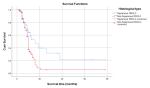
Survival analysis: in this cohort of 202 patients with non-small cell lung cancer, the total observation time was 1,664 person-months. The overall mortality rate was 5.05 deaths per 100 person-months of observation. The median survival time was 10 months, with an interquartile range of 7 to 16 months. Kaplan-Meier survival analysis was conducted to evaluate overall survival differences among subgroups of patients with non-small cell lung cancer. Patients with squamous cell carcinoma had significantly better survival outcomes than those with non-squamous cell carcinoma (median OS: 16 vs. 10 months). This difference was statistically significant (Log-Rank test: X² = 5.442, p = 0.020) (Figure 1). Furthermore, significant survival variation was observed across histological differentiation groups (Log-Rank: Χ² = 13.41, p = 0.0012).
Bivariate and multivariate analysis of the predictors of the overall survival of NSCLC patients: in bivariate Cox proportional hazards analysis, several variables were significantly associated with overall survival among NSCLC patients. Variables with p-values <0.25 in bivariate Cox proportional hazards analysis were entered into the multivariable Cox regression model. These variables included age, tumour location, tumour size, pleural effusion, primary tumour stage, nodal status, distant metastasis, histological subtype, histological differentiation, and performance status. In multivariate Cox proportional hazards analysis, age, primary tumour stage (T), regional lymph node involvement (N), presence of distant metastasis (M), histological subtype, histological differentiation, and performance status were significant predictors of survival among NSCLC patients (Table 5).
In this study, age was significantly associated with overall survival among NSCLC patients. Patients aged 40-59 years and? 60 years had significantly lower hazards of death compared with patients aged under 40 years (AHR = 0.22, 95% CI: 0.11-0.45 and AHR = 0.33, 95% CI: 0.17-0.65, respectively). Regarding TNM classification, patients with advanced primary tumour stages (T3-T4) had around five times higher risk of death than those with early-stage tumours (T1-T2) (AHR = 5.07, 95% CI: 1.08-23.88). Ipsilateral nodal disease did not independently affect survival. However, contralateral nodal involvement showed borderline statistical significance (AHR = 1.89, 95% CI: 1.00-3.59). Patients with distant metastasis (M1) had around three times higher hazard of death than non-metastatic patients (M0) (AHR = 3.14, 95% CI: 1.35-7.29) (Table 5). In this study, patients with non-squamous cell carcinoma had nearly twice the hazard of death compared with those with squamous cell carcinoma (AHR = 1.99; 95% CI: 1.09-3.60). Moderately differentiated tumours (AHR = 2.91; 95% CI: 1.34-6.32) and poorly differentiated/undifferentiated tumours (AHR = 3.35; 95% CI: 1.59-7.17) had progressively worse outcomes than well-differentiated tumours. Patients with poor performance status had more than a two-fold increased hazard of death compared with those with good performance status (AHR = 2.11; 95% CI: 1.15-3.90; p = 0.016) (Table 5).
In the present cohort, the overall mortality rate was 5.05 deaths per 100 person-months of observation, and the median survival time was 10 months. These findings align with published evidence in advanced lung cancer populations. In a large cohort of stage IV NSCLC patients, the median survival was reported as 10.0 months, with low one- and five-year survival rates indicating a high mortality burden in this disease context [17]. A Canadian population-based real-world analysis similarly reported median NSCLC survival of 10-13.7 months in one arm and 7.8 to 9.4 months in the other arm, although the treatment era modifies outcomes [18]. Population-based data from Shanghai also showed a median overall survival of 22.7 months overall, including earlier-stage disease; survival declines substantially with more advanced stages [19].
Meta-analytic evidence pooled from seven cohort studies and fifteen randomised controlled trials indicates that non-small cell lung cancer (NSCLC) in the absence of anticancer therapy is characterised by an exceptionally rapid mortality rate, estimated at 0.97, with a pooled mean survival of approximately 7.15 months [20]. In a French cohort spanning 2000-2020, survival outcomes for patients with metastatic lung cancer showed significant improvement over the past 20 years, with a reported median overall survival of 16.2 months [21]. In a nationwide retrospective cohort study evaluating the impact of pembrolizumab treatment duration and prognostic factors in advanced non-small cell lung cancer, the median overall survival following initiation of first-line pembrolizumab was 15.7 months [22]. These findings are higher than those observed in our study.
Patients aged 40-59 years and ≥ 60 years had significantly lower hazards of death compared with patients aged under 40 years. This observation is consistent with published articles. A study on the association between younger age and targetable genomic alterations and prognosis in non-small-cell lung cancer revealed that the survival of young patients with NCSLC is unexpectedly poor compared with other age groups, suggesting more aggressive disease biology [23]. Similarly, a retrospective study comparing survival outcomes in young NSCLC patients reported that patients younger than 40 years had a significantly worse prognosis than older patients [24]. Despite similar stages and tumour characteristics, patients younger than 45 years of age with non-small cell lung cancer in another study have a significantly worse prognosis than older patients [25]. This could be a result of the late presentation of young patients at the advanced stage during diagnosis [26].
In contrast, population-based analyses suggest improved lung cancer-specific survival compared with slightly older young adults, likely due to biological and treatment-related factors [27]. In the current cohort, advanced tumour characteristics defined by the TNM classification were strongly associated with survival, consistent with published evidence. Patients with advanced primary tumour stages (T3-T4) had a markedly higher mortality risk than those with early-stage tumours (T1-T2). This aligns with large population studies demonstrating that a higher T category correlates with progressively worse overall survival in lung cancer, and that survival declines markedly as the tumour extends locally and invades adjacent structures [28]. A large SEER database study of postoperative stage IIIA-N2 NSCLC patients also found that increasing tumour size groups were independently associated with worse survival [29]. T-stage information, including tumour size and extent of invasion, was also correlated with the prognosis of patients with NSCLC [30,31]. In the present cohort, ipsilateral nodal involvement did not independently predict mortality, whereas contralateral nodal disease showed borderline statistical significance. This pattern aligns with prior studies[32,33] that identify multiple metastatic nodal stations and lymph node ratio as prognostic factors. In contrast, contralateral nodal disease (N3) is associated with poorer outcomes in other cohort studies [34,35].
Patients with distant metastasis (M1) had a hazard of death approximately threefold higher than that of non-metastatic patients (M0). In line with this finding, large registry-based studies of NSCLC cohorts have consistently documented markedly worse outcomes for patients with metastatic disease compared with those without distant spread [36,37]. SEER database studies over the past few years have consistently shown that distant metastasis is associated with markedly shorter survival than in non-metastatic NSCLC, and that survival declines further with multi-site metastatic involvement [36].
This study also assessed the potential prognostic role of histology in NSCLC. Histology was found to be prognostic for survival, with better outcomes associated with squamous cell carcinoma and poorer outcomes with non-squamous cell carcinoma. The finding is supported by published research, although the direction varies by stage, treatment context, and patient population [13,38,39]. Evidence from large randomised trials confirms that squamous cell carcinoma is associated with superior survival compared with adenocarcinoma, even after controlling for clinical confounders [14]. A literature review of the prognostic and predictive role of histology in advanced NSCLC found that in half the articles, squamous cell carcinoma (or nonadenocarcinoma) was associated with better outcomes, while the other half found adenocarcinoma or nonsquamous histology associated with better outcomes [40].
In another study, postoperative adjuvant chemotherapy significantly improved overall and treatment-free survival in squamous cell carcinoma but not adenocarcinoma, highlighting differential chemosensitivity by histologic subtype [41]. In a French cohort of 6,101 patients, although overall five-year survival rates were poor; there were statistically significant differences in survival metrics when stratified by subtype, with squamous cell carcinoma showing slightly higher survival compared with adenocarcinoma and large-cell carcinoma [15].In another study, postoperative adjuvant chemotherapy significantly improved overall and treatment-free survival in squamous cell carcinoma but not in adenocarcinoma, highlighting differential chemosensitivity by histological subtype [41]. In a French cohort of 6,101 patients, although overall five-year survival rates were poor, there were statistically significant differences in survival metrics when stratified by subtype, with squamous cell carcinoma showing slightly higher survival than adenocarcinoma and large-cell carcinoma [15].
In the present study, histological differentiation was significantly associated with overall survival among patients with non-small cell lung cancer. Moderately differentiated and poorly differentiated/undifferentiated tumours were associated with progressively worse survival than well-differentiated tumours. A multicentre study of NSCLC cases found that moderately differentiated and undifferentiated/poorly differentiated tumours had a significantly higher risk of mortality than well-differentiated tumours [42]. In another study, poor differentiation was an independent prognostic factor after surgical resection [43]. Additionally, a retrospective cohort study of 531 patients found that histological grade was an independent prognostic factor in patients resected for all stages of NSCLC [12]. A recent National Cancer Database analysis also revealed that higher grade was associated with poorer overall survival in adenocarcinoma, although not in squamous cell carcinoma [44]. Poorly differentiated tumours consist of immature, aggressive cells that tend to grow more quickly, spread more often, and have a worse prognosis than tumours with well-differentiated cancer cells [45].
Patients with poor performance status had more than a two-fold increased hazard of death compared with those with good performance status in this study. Similarly, patients with NSCLC with poor performance status treated with immune checkpoint inhibitors had significantly worse survival outcomes and were significantly more likely to use health care services than those with favourable ECOG performance status. A meta-analysis of 19 studies further demonstrated that a poor performance status was significantly associated with worse overall survival compared with a favourable performance status. In addition, a prospective cohort study of patients with advanced NSCLC also identified poor ECOG performance status as an independent risk factor for poorer overall survival [46-48]. This could be a result of limited physical activity and impaired ability to tolerate intensive therapies.
Limitations: first, the retrospective design may be subject to incomplete documentation, missing variables, and misclassification of clinical, imaging, and histopathological data. Exclusion of patients with incomplete medical records may have introduced selection bias, particularly if excluded patients differed systematically from included participants.
In the present cohort, the overall incidence rate of death was 5.05 deaths per 100 person-months of follow-up, and the median survival time was 10 months. Old age, higher primary tumour stage (T), contralateral regional lymph node involvement (N), presence of distant metastasis (M), non-squamous histological subtype, high-grade tumour, and poor performance status were found to be significant predictors of survival among NSCLC patients.
What is known about this topic
- Although the tumour-node-metastasis (TNM) staging system remains the gold standard for prognostication, it does not fully explain survival variability among NSCLC patients;
- Histological subtype and tumour differentiation have been increasingly recognised as important prognostic indicators;
- However, findings remain inconsistent across populations and treatment settings, and most evidence is derived from high-income countries where molecular diagnostics and targeted therapies are widely available.
What this study adds
- We found a median survival of 10 months and a high proportion of patients (73.8%) presenting with advanced disease;
- We identified old age, advanced primary tumour stage (T3-T4), distant metastasis, contralateral nodal involvement, non-squamous histological subtype, high-grade tumour and poor performance status as independent predictors of mortality among NSCLC patients;
- Histological subtype and tumour differentiation provide prognostic information beyond the diagnosis of NSCLC and are valuable for risk stratification and guiding treatment decision-making in settings where access to advanced molecular testing is limited.
The authors declare no competing interests.
Binalfew Tsehay: conceptualisation, data acquisition, analysis, interpretation, and writing the manuscript. Mekbeb Afework: conceptualisation, reviewing the proposal, the manuscript, and data interpretation. Wondwossen Ergete: conceptualisation, reviewing proposal and manuscript. Yonas Dandena: reviewing the manuscript and data interpretation. All the authors read and approved the final version of this manuscript.
Table 1: sociodemographic and clinical characteristics of patients with NSCLC (n = 202)
Table 2: baseline computerised tomography imaging features of NSCLC patients (n = 202)
Table 3: histological subtypes and histological differentiation of NSCLC patients (n = 202)
Table 4: tumour stage, performance status, and treatment patterns of NSCLC patients (n = 202)
Table 5: bivariate and multivariate analysis of the predictors on the overall survival of NSCLC patients
Figure 1: Kaplan-Meier survival curves by histological subtype in non-small cell lung cancer patients
- Hendriks LE, Remon J, Faivre-Finn C, Garassino MC, Heymach JV, Kerr KM et al. Non-small-cell lung cancer. Nat Rev Dis Primers. 2024 Sep 26;10(1):71. PubMed | Google Scholar
- Thandra KC, Barsouk A, Saginala K, Aluru JS, Barsouk A. Epidemiology of lung cancer. Contemp Oncol (Pozn). 2021;25(1):45-52. PubMed | Google Scholar
- Leiter A, Veluswamy RR, Wisnivesky JP. The global burden of lung cancer: current status and future trends. Nat Rev Clin Oncol. 2023 Sep;20(9):624-639. PubMed | Google Scholar
- Travis WD, Brambilla E, Burke AP, Marx A, Nicholson AG. Introduction to The 2015 World Health Organization Classification of Tumors of the Lung, Pleura, Thymus, and Heart. J Thorac Oncol. 2015 Sep;10(9):1240-1242. PubMed | Google Scholar
- Thomas A, Liu SV, Subramaniam DS, Giaccone G. Refining the treatment of NSCLC according to histological and molecular subtypes. Nat Rev Clin Oncol. 2015 Sep;12(9):511-26. PubMed | Google Scholar
- Rodak O, Peris-Díaz MD, Olbromski M, Podhorska-Okolów M, Dziegiel P. Current Landscape of Non-Small Cell Lung Cancer: Epidemiology, Histological Classification, Targeted Therapies, and Immunotherapy. Cancers (Basel). 2021 Sep 20;13(18):4705. PubMed | Google Scholar
- Cuesta JCP, Aragón CJ, Franch NM, Navarro JAC, de Aguiar Quevedo K, Martínez MA et al. Prognostic Factors in Non-Small Cell Lung Cancer Less Than 3 Centimeters: Actuarial Analysis, Accumulative Incidence and Risk Groups. Arch Bronconeumol. 2015 Sep;51(9):431-9. English, Spanish. PubMed | Google Scholar
- Jazieh AR, Hussain M, Howington JA, Spencer H, Husain M, Grismer JT et al. Prognostic factors in patients with surgically resected stages I and II non-small cell lung cancer. Ann Thorac Surg. 2000 Oct;70(4):1168-71. PubMed | Google Scholar
- Maeda R, Yoshida J, Ishii G, Hishida T, Aokage K, Nishimura M et al. Long-term survival and risk factors for recurrence in stage I non-small cell lung cancer patients with tumors up to 3 cm in maximum dimension. Chest. 2010 Aug;138(2):357-62. PubMed | Google Scholar
- Kratz JR, He J, Van Den Eeden SK, Zhu Z-H, Gao W, Pham PT et al. A practical molecular assay to predict survival in resected non-squamous, non-small-cell lung cancer: development and international validation studies. Lancet. 2012 Mar 3;379(9818):823-32. PubMed | Google Scholar
- Padilla J, Calvo V, Penalver J, Zarza A G, Pastor J, Blasco E et al. Survival and risk model for stage IB non-small cell lung cancer. Lung Cancer. 2002 Apr;36(1):43-8. PubMed | Google Scholar
- Yasukawa M, Sawabata N, Kawaguchi T, Kawai N, Nakai T, Ohbayashi C et al. Histological Grade: Analysis of Prognosis of Non-small Cell Lung Cancer After Complete Resection. In Vivo. 2018 Nov-Dec;32(6):1505-1512. PubMed | Google Scholar
- Khan OA, Fitzgerald JJ, Field ML, Soomro I, Beggs FD, Morgan WE et al. Histological determinants of survival in completely resected T1-2N1M0 nonsmall cell cancer of the lung. Ann Thorac Surg. 2004 Apr;77(4):1173-8. PubMed | Google Scholar
- Scagliotti GV, De Marinis F, Rinaldi M, Crino L, Gridelli C, Ricci S et al. The role of histology with common first-line regimens for advanced non-small cell lung cancer: a brief report of the retrospective analysis of a three-arm randomized trial. J Thorac Oncol. 2009 Dec;4(12):1568-71. PubMed | Google Scholar
- Molinier O, Goupil F, Debieuvre D, Auliac J-B, Jeandeau S, Lacroix S et al. Five-year survival and prognostic factors according to histology in 6101 non-small-cell lung cancer patients. Respir Med Res. 2020 Mar;77:46-54. PubMed | Google Scholar
- Trani L, Myerson J, Ashley S, Young K, Sheri A, Hubner R et al. Histology classification is not a predictor of clinical outcomes in advanced non-small cell lung cancer (NSCLC) treated with vinorelbine or gemcitabine combinations. Lung Cancer. 2010 Nov;70(2):200-4. PubMed | Google Scholar
- Peng H, Ma M, Han B. [Survival analysis of 1,742 patients with stage IV non-small cell lung cancer]. Zhongguo Fei Ai Za Zhi. 2011 Apr;14(4):362-6. Chinese. PubMed | Google Scholar
- Carroll R, Bortolini M, Calleja A, Munro R, Kong S, Daumont MJ et al. Trends in treatment patterns and survival outcomes in advanced non-small cell lung cancer: a Canadian population-based real-world analysis. BMC Cancer. 2022 Mar 10;22(1):255. PubMed | Google Scholar
- Fan H, Shao Z-Y, Xiao Y-Y, Xie Z-H, Chen W, Xie H et al. Incidence and survival of non-small cell lung cancer in Shanghai: a population-based cohort study. BMJ Open. 2015 Dec 23;5(12):e009419. PubMed | Google Scholar
- Wao H, Mhaskar R, Kumar A, Miladinovic B, Djulbegovic B. Survival of patients with non-small cell lung cancer without treatment: a systematic review and meta-analysis. Syst Rev. 2013 Feb 4;2:10. PubMed | Google Scholar
- Basse C, Carton M, Milder M, Geiss R, Du Rusquec P, Daniel C et al. Real-World Survival Impact of New Treatment Strategies for Lung Cancer: A 2000-2020 French Cohort. Cancers (Basel). 2024 Aug 5;16(15):2768. PubMed | Google Scholar
- Rousseau A, Michiels S, Simon-Tillaux N, Lolivier A, Bonastre J, Planchard D et al. Impact of pembrolizumab treatment duration on overall survival and prognostic factors in advanced non-small cell lung cancer: a nationwide retrospective cohort study. Lancet Reg Health Eur. 2024 Jun 29;43:100970. PubMed | Google Scholar
- Sacher AG, Dahlberg SE, Heng J, Mach S, Jänne PA, Oxnard GR. Association Between Younger Age and Targetable Genomic Alterations and Prognosis in Non-Small-Cell Lung Cancer. JAMA Oncol. 2016 Mar;2(3):313-20. PubMed | Google Scholar
- Bratova M, Brat K, Hurdalkova K, Barinova M, Drosslerova M, Kultan J et al. Lung cancer versus “young cancer”: is non-small cell lung cancer in young patients a different entity? Journal of adolescent and young adult oncology. 2022 Oct;11(5):451-458. PubMed | Google Scholar
- Bryant AS, Cerfolio RJ. Differences in outcomes between younger and older patients with non-small cell lung cancer. Ann Thorac Surg. 2008 May;85(5):1735-9; discussion 1739. PubMed | Google Scholar
- Hughes DJ, Kapiris M, Podvez Nevajda A, McGrath H, Stavraka C, Ahmad S et al. Non-small cell lung cancer (NSCLC) in young adults, age< 50, is associated with late stage at presentation and a very poor prognosis in patients who do not have a targeted therapy option: a real-world study. Cancers. 2022 Dec 9;14(24):6056. PubMed | Google Scholar
- Dingillo G, Bassiri A, Badrinathan A, Alvarado CE, Sinopoli J, Tapias L et al. Lung Cancer in Young Patients is Associated With More Advanced Disease but Better Overall Survival. J Surg Res. 2023 Dec;292:307-316. PubMed | Google Scholar
- Lee J, Lee J, Hong YS, Lee G, Park J, Jeon YJ et al. Impact of tumor size by clinical N subclassification and histology in trimodality-treated N2 non-small cell lung cancer. Sci Rep. 2025 May 17;15(1):17195. PubMed | Google Scholar
- Zhang K, Cai J, Lu C, Zhu Q, Zhan C, Shen Y et al. Tumor size as a predictor for prognosis of patients with surgical IIIA-N2 non-small cell lung cancer after surgery. J Thorac Dis. 2021 Jul;13(7):4114-4124. PubMed | Google Scholar
- Mao Q, Xia W, Dong G, Chen S, Wang A, Jin G et al. A nomogram to predict the survival of stage IIIA-N2 non-small cell lung cancer after surgery. J Thorac Cardiovasc Surg. 2018 Apr;155(4):1784-1792.e3. PubMed | Google Scholar
- Chen C-Y, Wu B-R, Chen C-H, Cheng W-C, Chen W-C, Liao W-C et al. Prognostic Value of Tumor Size in Resected Stage IIIA-N2 Non-Small-Cell Lung Cancer. J Clin Med. 2020 May 1;9(5):1307. PubMed | Google Scholar
- Kang CH, Ra YJ, Kim YT, Jheon S-H, Sung S-W, Kim JH. The impact of multiple metastatic nodal stations on survival in patients with resectable N1 and N2 nonsmall-cell lung cancer. Ann Thorac Surg. 2008 Oct;86(4):1092-7. PubMed | Google Scholar
- Luo B, Liang S, Zhong L, Gao W, Zeng M, Chen Y et al. Impact of N1 lymph node burden on survival outcomes in non-skip pN2 non-small-cell lung cancer. Lung Cancer. 2025 Dec;210:108839. PubMed | Google Scholar
- Gopal R, Starkschall G, Tucker SL, Cox JD, Liao Z, Hanus M et al. Effects of radiotherapy and chemotherapy on lung function in patients with non-small-cell lung cancer. Int J Radiat Oncol Biol Phys. 2003 May 1;56(1):114-20. PubMed | Google Scholar
- Morris ZS, Cannon DM, Morris BA, Bentzen SM, Kozak KR. Impact of a Contralateral Tumor Nodule on Survival in Non-Small-Cell Lung Cancer. J Thorac Oncol. 2015 Nov;10(11):1608-15. PubMed | Google Scholar
- Wu B, Wei S, Tian J, Song X, Hu P, Cui Y. [Comparison of the Survival Time in the Non-small Cell Lung Cancer Patients with Different Organ Metastasis]. Zhongguo Fei Ai Za Zhi. 2019 Feb 20;22(2):105-110. Chinese. PubMed | Google Scholar
- Bertram S, Schliemann A, Katalinic A, Kim-Wanner S-Z, Pritzkuleit R, Twardella D et al. Overall survival and progression-free survival of patients with non-small cell lung cancer and either limited or extensive synchronous metastatic spread in Germany-a population-based cancer registry cohort study. Transl Lung Cancer Res. 2025 Oct 31;14(10):4436-4448. PubMed | Google Scholar
- Grosu HB, Manzanera A, Shivakumar S, Sun S, Noguras Gonzalez G, Ost DE. Survival disparities following surgery among patients with different histological types of non-small cell lung cancer. Lung Cancer. 2020 Feb;140:55-58. PubMed | Google Scholar
- Guo S, Reddy CA, Chao ST, Suh JH, Barnett GH, Vogelbaum MA et al. Impact of non-small cell lung cancer histology on survival predicted from the graded prognostic assessment for patients with brain metastases. Lung Cancer. 2012 Aug;77(2):389-93. PubMed | Google Scholar
- Hirsch FR, Spreafico A, Novello S, Wood MD, Simms L, Papotti M. The prognostic and predictive role of histology in advanced non-small cell lung cancer: a literature review. J Thorac Oncol. 2008 Dec;3(12):1468-81. PubMed | Google Scholar
- Hsiao SH, Chen WT, Chung CL, Chou YT, Lin SE, Hong SY et al. Comparative survival analysis of platinum-based adjuvant chemotherapy for early-stage squamous cell carcinoma and adenocarcinoma of the lung. Cancer Med. 2022 May;11(10):2067-2078. PubMed | Google Scholar
- Sun Z, Aubry M-C, Deschamps C, Marks RS, Okuno SH, Williams BA et al. Histologic grade is an independent prognostic factor for survival in non-small cell lung cancer: an analysis of 5018 hospital- and 712 population-based cases. J Thorac Cardiovasc Surg. 2006 May;131(5):1014-20. PubMed | Google Scholar
- Liu CH, Peng YJ, Wang HH, Chen YC, Tsai CL, Chian CF et al. Heterogeneous prognosis and adjuvant chemotherapy in pathological stage I non-small cell lung cancer patients. Thorac Cancer. 2015 Sep;6(5):620-8. PubMed | Google Scholar
- Endo T, Crane J, Trivedi J, Black M, Fox M, van Berkel V. Does grade matter? Evaluation of histologic grade in patients with stage 1 non-small cell lung cancer using the National Cancer Database. JTCVS Open. 2025 Mar 11;25:393-405. PubMed | Google Scholar
- Canadian Cancer Society. Grading cancer. 2025. Accessed on 2026 January 02.
- Meyers DE, Pasternak M, Dolter S, Grosjean HA, Lim CA, Stukalin I et al. Impact of Performance Status on Survival Outcomes and Health Care Utilization in Patients With Advanced NSCLC Treated With Immune Checkpoint Inhibitors. JTO Clin Res Rep. 2023 Feb 24;4(4):100482. PubMed | Google Scholar
- Dall'Olio FG, Maggio I, Massucci M, Mollica V, Fragomeno B, Ardizzoni A. ECOG performance status =2 as a prognostic factor in patients with advanced non small cell lung cancer treated with immune checkpoint inhibitors-A systematic review and meta-analysis of real world data. Lung Cancer. 2020 Jul;145:95-104. PubMed | Google Scholar
- Sehgal K, Gill RR, Widick P, Bindal P, McDonald DC, Shea M et al. Association of Performance Status With Survival in Patients With Advanced Non-Small Cell Lung Cancer Treated With Pembrolizumab Monotherapy. JAMA Netw Open. 2021 Feb 1;4(2):e2037120. PubMed | Google Scholar
Search
This article authors
On Pubmed
On Google Scholar
Citation [Download]
Navigate this article
Similar articles in
Key words
Tables and figures

Article metrics
Recently from the PAMJ



Authors´ services
