Diagnostic challenge of a pelvic cystic mass: localized pseudomyxoma peritonei secondary to a ruptured low-grade appendiceal mucinous neoplasm
Salim Ben Souayah, Hajar Khaoulani, Youssef Bouktib, Ayoub El Hajjami, Badr Boutakioute, Meriem Ouali Idrissi, Najat Cherif Idrissi El Gannouni
Corresponding author: Salim Ben Souayah, Radiology Department, Ar-Razi Hospital, University Hospital Center Mohammed VI, Marrakech, Morocco 
Received: 18 Nov 2025 - Accepted: 06 Aug 2026 - Published: 27 Oct 2026
Domain: Radiology
Keywords: Pseudomyxoma peritonei, mucinous tumor, low-grade appendiceal mucinous neoplasms, magnetic resonance imaging, case report
Funding: This work received no specific grant from any funding agency in the public, commercial, or non-profit sectors.
©Salim Ben Souayah et al. Pan African Medical Journal (ISSN: 1937-8688). This is an Open Access article distributed under the terms of the Creative Commons Attribution International 4.0 License (https://creativecommons.org/licenses/by/4.0/), which permits unrestricted use, distribution, and reproduction in any medium, provided the original work is properly cited.
Cite this article: Salim Ben Souayah et al. Diagnostic challenge of a pelvic cystic mass: localized pseudomyxoma peritonei secondary to a ruptured low-grade appendiceal mucinous neoplasm. Pan African Medical Journal. 2026;55:97. [doi: 10.11604/pamj.2026.55.97.50274]
Available online at: https://www.panafrican-med-journal.com//content/article/55/97/full
 PDF in process
Case report 

Diagnostic challenge of a pelvic cystic mass: localized pseudomyxoma peritonei secondary to a ruptured low-grade appendiceal mucinous neoplasm
Diagnostic challenge of a pelvic cystic mass: localized pseudomyxoma peritonei secondary to a ruptured low-grade appendiceal mucinous neoplasm
 Salim Ben Souayah1,2,&, Hajar Khaoulani1,2, Youssef Bouktib1,2, Ayoub El Hajjami1,2, Badr Boutakioute1,2, Meriem Ouali Idrissi1,2, Najat Cherif Idrissi El Gannouni1,2
&Corresponding author
Pseudomyxoma peritonei (PMP) is a rare condition that usually results from the rupture of a low-grade appendiceal mucinous neoplasm (LAMN). Its diagnosis may be challenging when imaging cannot clearly identify the primary site of the lesion. We report the case of an 86-year-old woman who presented with chronic abdominal and pelvic pain associated with weight loss and asthenia. Contrast-enhanced computed tomography (CT) and magnetic resonance imaging (MRI) showed a predominantly cystic right pelvic mass of uncertain appendiceal or ovarian origin, associated with pancreatic and hepatic cystic lesions suspicious for secondary involvement. The patient underwent cytoreductive surgery with right hemicolectomy and right adnexectomy. Histopathological examination confirmed a ruptured LAMN complicated by localized pseudomyxoma peritonei, while the right ovary and fallopian tube were free of tumor. This case highlights the diagnostic challenges of pelvic mucinous tumors and the complementary role of imaging and histopathology in establishing the final diagnosis.

Pseudomyxoma peritonei (PMP) is a rare clinicopathological condition characterized by progressive accumulation of mucinous ascites associated with dissemination of mucin-producing epithelial cells within the peritoneal cavity [1]. In most cases, it originates from the rupture of a low-grade appendiceal mucinous neoplasm (LAMN), whereas primary mucinous tumors arising from other organs are uncommon [1].
Because of its nonspecific clinical presentation and variable imaging findings, PMP remains a diagnostic challenge. Patients commonly present with chronic abdominal pain, abdominal distension or, less frequently, bowel obstruction, while the diagnosis is often established only after surgery and histopathological examination [1,2]. Early recognition is important because it helps guide appropriate surgical management [1].
Contrast-enhanced computed tomography (CT) is the imaging modality of choice for the initial evaluation of PMP, whereas magnetic resonance imaging (MRI) provides complementary information, particularly for characterizing indeterminate pelvic lesions and assessing their relationship with adjacent organs [2,3]. However, determining the exact origin of a mucinous pelvic mass may remain difficult when the appendix and the right ovary cannot be clearly distinguished on imaging. In such cases, histopathological examination remains essential for establishing the final diagnosis [4].
We report the case of an elderly woman with localized PMP secondary to a ruptured LAMN that initially presented as a pelvic cystic mass of uncertain origin. This case highlights the diagnostic challenge of distinguishing an appendiceal mucinous neoplasm from an ovarian mucinous tumor and emphasizes the complementary roles of CT, MRI, and histopathological examination in establishing the final diagnosis.

Patient information: 86-year-old woman, gravida 8 para 8, with a history of hypertension complicated by hypertensive heart disease treated with antiplatelet therapy and dyslipidemia treated with statins, with no previous abdominal surgery.
Clinical findings: the patient presented with a six-month history of progressive abdominal and pelvic pain, predominantly in the hypogastric region, associated with asthenia, anorexia, and weight loss. On admission, she was hemodynamically stable. Abdominal examination revealed a soft abdomen with tenderness over the hypogastric region and both iliac fossae. No palpable abdominal mass, hernia, or peripheral lymphadenopathy was noted. The remainder of the physical examination was unremarkable.
Timeline of current episode: 6 months before admission, the patient developed progressive diffuse abdominal pain, predominantly in the hypogastric region, associated with asthenia, anorexia, and weight loss. At admission, clinical examination, laboratory investigations and contrast-enhanced CT were performed. Later on, pelvic MRI was requested for further characterization of the lesion. The patient then underwent cytoreductive surgery including peritonectomy, right hemicolectomy and right adnexectomy. Histopathological examination confirmed a ruptured low-grade appendiceal mucinous neoplasm complicated by localized pseudomyxoma peritonei. At the last follow-up visit, the patient reported marked improvement in abdominal pain despite mild persistent abdominal discomfort. She was subsequently lost to follow-up, and no postoperative imaging was available.
Diagnostic assessment: laboratory investigations revealed elevated serum CA-125 (56.20 U/mL) and CA 19-9 (43.59 U/mL). Contrast-enhanced computed tomography (CT) demonstrated a predominantly cystic right latero-uterine pelvic mass closely related to the uterus and ileocecal region, without clear visualization of either the appendix or the right ovary (Figure 1). Two additional cystic lesions involving the liver and pancreas showed imaging characteristics similar to the pelvic lesion and were considered suspicious for possible secondary involvement (Figure 2).
Pelvic magnetic resonance imaging confirmed a predominantly cystic right pelvic mass containing thin internal vegetations (Figure 3). However, the origin of the lesion remained uncertain because neither the appendix nor the right ovary could be identified separately. Magnetic resonance imaging also showed diffuse endometrial thickening suggestive of endometrial hyperplasia. Based on the imaging findings, the main differential diagnoses included an appendiceal mucinous neoplasm and a primary ovarian mucinous tumor.
Diagnosis: the patient underwent cytoreductive surgery including peritonectomy, right hemicolectomy, and right adnexectomy (Figure 4 and Figure 5). Histopathological examination confirmed a ruptured low-grade appendiceal mucinous neoplasm complicated by localized pseudomyxoma peritonei. The right ovary and fallopian tube were free of tumor involvement, confirming the appendiceal origin of the disease (Figure 6).
Therapeutic interventions: considering the intraoperative findings, cytoreductive surgery consisting of peritonectomy, right hemicolectomy, and right adnexectomy was performed. Hyperthermic intraperitoneal chemotherapy (HIPEC) was not performed because it was unavailable at our institution.
Follow-up and outcome of interventions: the postoperative course was uneventful. At the last follow-up visit, the patient reported a marked improvement in abdominal pain, although mild abdominal discomfort persisted. No postoperative complications were observed. The patient was subsequently lost to follow-up; therefore, no postoperative imaging or long-term clinical outcome was available.
Patient perspective: "since the operation, my abdominal pain has significantly improved. Although I still have mild discomfort, I feel much better than before surgery, and I am overall satisfied with the treatment I received."
Informed consent: written informed consent for publication of this case report and the accompanying images was obtained from the patient's legal caregivers.
Pseudomyxoma peritonei (PMP) is a rare clinicopathological condition characterized by progressive accumulation of mucinous ascites associated with dissemination of mucin-producing epithelial cells within the peritoneal cavity. In most cases, it originates from a ruptured low-grade appendiceal mucinous neoplasm (LAMN), whereas other primary sites are uncommon [1,2].
Preoperative diagnosis remains challenging because both the clinical presentation and imaging findings are often nonspecific. In women, differentiating an appendiceal mucinous neoplasm from a primary ovarian mucinous tumor may be particularly difficult when the appendix and ovary cannot be clearly identified separately, as illustrated in our patient. This distinction is clinically important because ovarian involvement is frequently secondary to an appendiceal primary tumor rather than a primary ovarian neoplasm [3,4].
Contrast-enhanced CT is considered the imaging modality of choice for the initial evaluation of PMP because it accurately demonstrates the primary lesion, the extent of peritoneal disease, mucinous ascites, peritoneal implants, and scalloping of the visceral surfaces [3,5]. Magnetic resonance imaging provides complementary information, particularly for characterizing indeterminate pelvic lesions because of its superior soft-tissue contrast [3]. The imaging appearance of appendiceal mucoceles has been well described, although identifying the exact site of origin may remain difficult when adjacent pelvic organs are closely related [6]. Despite the combined use of CT and MRI, the origin of the pelvic mass could not be confidently established before surgery in our patient. Another noteworthy finding was the presence of pancreatic and hepatic cystic lesions showing imaging characteristics similar to those of the pelvic lesion. Although these findings raised the possibility of secondary involvement, their exact nature remains uncertain because neither histopathological confirmation nor postoperative imaging follow-up was available. They should therefore be interpreted cautiously [1].
Histopathological examination remains the gold standard for confirming the diagnosis and determining the primary site of disease [7]. In our case, examination of the surgical specimen confirmed a ruptured LAMN complicated by localized PMP, while the right ovary and fallopian tube were free of tumor involvement, confirming the appendiceal origin. The current PSOGI consensus has standardized the pathological classification and reporting of appendiceal mucinous neoplasms and PMP, improving diagnostic consistency and communication between pathologists and clinicians [8].
Complete cytoreductive surgery remains the cornerstone of treatment for PMP. In specialized centers, it is usually combined with hyperthermic intraperitoneal chemotherapy (HIPEC), which has been associated with improved long-term disease control in selected patients [9]. However, HIPEC was unavailable at our institution, and the patient therefore underwent cytoreductive surgery alone. Our case highlights the diagnostic challenge of determining the origin of a cystic pelvic mass when the appendix and ovary cannot be clearly distinguished on imaging. It also emphasizes the complementary roles of CT, MRI, surgery and histopathological examination in establishing the final diagnosis. Appendiceal mucinous neoplasms should therefore be considered in the differential diagnosis of complex cystic pelvic lesions, particularly in elderly women.
Pseudomyxoma peritonei should be considered in the differential diagnosis of cystic pelvic masses, particularly when the appendix cannot be clearly distinguished from the ovary on imaging. Although CT and MRI play a key role in preoperative assessment, histopathological examination remains essential for confirming the diagnosis and identifying the primary site.
The authors declare no competing interests.
All the authors have read and approved the final version of this manuscript.
Figure 1: A, B) abdomino-pelvic CECT showing a lateral right uterine mass in close contact with the uterus
Figure 2: A,B) abdomino-pelvic CECT - cystic lesion shown in the pancreatic body and segment II of the liver
Figure 3: A-E) MRI sequences showing a right pelvic mass, predominantly cystic, with thin sparse vegetations
Figure 4: intra-operative image showing the tumour mass attached to the appendix
Figure 5: image of the surgical specimen of the hemicolectomy and right anexectomy
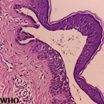
Figure 6: histopathological features of ruptured low-grade appendiceal mucinous neoplasm; A) cystic component; B) appendiceal perforation site (asterisk); HES staining, x200
- Shaib WL, Assi R, Shamseddine A, Alese OB, Staley C, Memis B et al. Appendiceal mucinous neoplasms: diagnosis and management. Oncologist. 2017;22(9):1107-16.. PubMed | Google Scholar
- Loungnarath R, Causeret S, Brigand C, Gilly FN, Glehen O. Pseudomyxoma peritonei: new concept and new therapeutic approach. Ann Chir. 2005;130(2):63-9. PubMed | Google Scholar
- Tirumani SH, Fraser-Hill M, Auer R, Shabana W, Walsh C, Lee F et al. Mucinous neoplasms of the appendix: a current comprehensive clinicopathologic and imaging review. Cancer Imaging. 2013;13(1):14-25. PubMed | Google Scholar
- Spyropoulos C, Rentis A, Alexakis E, Triantafillidis JK, Vagianos C. Appendiceal mucocele and pseudomyxoma peritonei: the clinical boundaries of a subtle disease. Am J Case Rep. 2014;15:355-60. PubMed | Google Scholar
- Pickhardt PJ, Levy AD, Rohrmann CA Jr, Kende AI. Primary neoplasms of the appendix: radiologic spectrum of disease with pathologic correlation. Radiographics. 2003;23(3):645-62. PubMed | Google Scholar
- Madwed D, Mindelzun R, Jeffrey RB Jr. Mucocele of the appendix: imaging findings. AJR Am J Roentgenol. 1992;159(1):69-72. PubMed | Google Scholar
- Pai RK, Longacre TA. Appendiceal mucinous neoplasms: clinicopathologic study of 116 cases with analysis of factors predicting recurrence. Am J Surg Pathol. 2009;33(10):1425-39. PubMed | Google Scholar
- Carr NJ, Cecil TD, Mohamed F, Sobin LH, Sugarbaker PH, González-Moreno S et al. A consensus for classification and pathologic reporting of pseudomyxoma peritonei and associated appendiceal neoplasia: the results of the Peritoneal Surface Oncology Group International (PSOGI) modified Delphi process. Am J Surg Pathol. 2016;40(1):14-26. PubMed | Google Scholar
- González-Moreno S, Sugarbaker PH. Right hemicolectomy does not confer a survival advantage in patients with mucinous carcinoma of the appendix and peritoneal seeding. Br J Surg. 2004;91(3):304-11. PubMed | Google Scholar
Search
This article authors
On Pubmed
On Google Scholar
Citation [Download]
Navigate this article
Similar articles in
Key words
Tables and figures


Article metrics
Recently from the PAMJ





Authors´ services