
Retrorectal cystic hamartoma (tailgut cyst): a case report
Wael Boujelbène, Amina Chaka, Amine Zouari, Nizar Kardoun, Amine Chaabouni, Nermine Ellouze, Islem Bradai, Soumaya Graja, Salma Ketata, Tahya Boudawara, Salah Boujelbène
Corresponding author: Wael Boujelbène, General Surgery Department, Habib Bourguiba Hospital, Sfax, Tunisia 
Received: 28 Aug 2025 - Accepted: 03 Jul 2026 - Published: 23 Jul 2026
Domain: General surgery
Keywords: Retrorectal tumors, tailgut cyst, treatment, prognosis, case report
Funding: This work received no specific grant from any funding agency in the public, commercial, or non-profit sectors.
©Wael Boujelbène et al. Pan African Medical Journal (ISSN: 1937-8688). This is an Open Access article distributed under the terms of the Creative Commons Attribution International 4.0 License (https://creativecommons.org/licenses/by/4.0/), which permits unrestricted use, distribution, and reproduction in any medium, provided the original work is properly cited.
Cite this article: Wael Boujelbène et al. Retrorectal cystic hamartoma (tailgut cyst): a case report. Pan African Medical Journal. 2026;54:97. [doi: 10.11604/pamj.2026.54.97.49003]
Available online at: https://www.panafrican-med-journal.com//content/article/54/97/full
 PDF in process
 Case report 

Retrorectal cystic hamartoma (tailgut cyst): a case report
Retrorectal cystic hamartoma (tailgut cyst): a case report
 Wael Boujelbène1,&, Amina Chaka2, Amine Zouari1, Nizar Kardoun1, Amine Chaabouni1, Nermine Ellouze3, Islem Bradai4, Soumaya Graja3, Salma Ketata4, Tahya Boudawara3, Salah Boujelbène1
&Corresponding author
Retrorectal (presacral) tumors are rare lesions arising from embryonic hindgut remnants, with the tailgut cyst being the most common subtype. They are often asymptomatic, and malignant transformation has been reported but remains uncommon. We report a 58-year-old woman with four months of pelvic pain. Clinical examination and laboratory tests were unremarkable. Computed tomography (CT) scan and magnetic resonance imaging (MRI) revealed a tailgut cyst. A complete laparoscopic excision was performed. Histopathology confirmed a reworked tailgut cyst with no evidence of malignancy. This case highlights the importance of imaging for accurate diagnosis and supports complete surgical excision, preferably via a minimally invasive approach, as the definitive management to prevent recurrence and exclude malignancy.

Retrorectal tumors are rare lesions developed from a residue of the primitive embryonic hindgut, with an estimated incidence of 1 in 40,000 people [1]. The tailgut cyst or cystic hamartoma is the most common retrorectal tumor. It is common in middle-aged women but rarely in children [1]. These tumors are often asymptomatic but can present non-specific symptoms related to an increase in cyst size or complications such as infections [1]. Although most cysts are benign, 30% of cases reported in the literature were malignant [2]. The surgical treatment consists of a complete cyst resection [3]. We report a case of a symptomatic tailgut cyst in a 58-year-old woman, which was successfully managed via complete laparoscopic excision and is described herein.

Patient information: a 58-year-old female patient with a history of hypothyroidism under levothyroxine, postmenopausal, non-smoker, non alcoholic.
Clinical findings: clinical examination found a patient in good general condition, apyretic. On abdominal exam, we find a non-distended, soft, depressed, and painless abdomen without a palpable mass. The rectal examination reveals no palpable mass with normal-colored stool. The gynecological examination was without abnormality: The vaginal examination revealed soft and elastic walls. It was free of any masses or lesions. The cervix was centrally positioned, smooth, and mobile, with no signs of tenderness upon palpation. The uterus was of normal size, anteverted, and mobile, with no abnormalities detected. The clinical examination was thorough and included an evaluation of the lymph node regions, particularly the inguinal areas, which were found to be free of any abnormalities.
Timeline of current episode: the patient has been experiencing paroxysmal pelvic pain for the past four months, without any associated bowel disorders or alterations in general health. There have been no significant changes in weight, energy levels, or appetite, and the patient does not report any systemic symptoms such as fever or excessive fatigue. The pain remains intermittent, with no abnormal uterine bleeding or other gynecological symptoms.
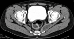
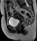
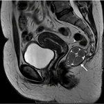
Diagnostic assessment: the biological assessment was without any particularity. There was no evidence of biological inflammatory syndrome or nutritional disorders. The tumor marker (CEA) was within normal limits. The abdominopelvic CT scan showed a hypodense fusiform thickening of the levator ani muscle on the left side of the pelvis, 5 cm long, with a liquid density, not enhanced after injection of iodinated contrast, with a nonspecific aspect, associated with a small calcified lymph node (Figure 1). The pelvic MRI showed a 42 x 37mm left paramedian mass, developed in the supra-levator space, with a hyper signal in T1 and an intermediate signal in T2, with diffusion restriction. This mass has thin partitions at the origin of a multilocular aspect and contains peripheral parietal macro calcification. It is close to the left-lateral wall of the rectum and leans against the homolateral iliococcygeus muscle without any sign of invasion of the integrity of the surrounding fat and the internal sphincter of the anus.
Diagnosis: the topography of the mass and the semiological aspect evoke a retrorectal tailgut cyst (Figure 2).
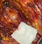
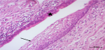
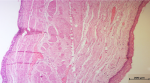
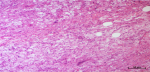
Therapeutic intervention: the patient underwent laparoscopic surgery, as this approach provides excellent exposure to major pelvic structures, which is crucial for the safe management of retrorectal lesions. Additionally, the minimally invasive nature of this technique offers significant advantages, including reduced postoperative pain and faster recovery compared to open surgery. In our case, the laparoscopic per-operative exploration revealed a retrorectal cystic mass measuring approximately 40 mm (Figure 3). The mesorectum was first dissected off the anterior portion of the lesion, then the lesion was separated from the presacral fascia. The dissection resulted in an accidental perforation of the cyst with the release of liquid content. The cyst was removed from the abdominal cavity. On macroscopic examination, the surgical specimen measured 50 x 50 x 25 mm. Sectioning revealed the presence of a multilocular cyst filled with a translucent substance. Histological analysis showed that an epithelium of keratinized squamous type, respiratory type, or regular mucin-secreting columnar type lined the cystic compartments. In some areas, this epithelium was destroyed and replaced by an inflammatory reaction characterized by macrophages and foreign-body-type giant cells. The cyst wall was fibromuscular and inflammatory. Importantly, no signs of malignancy were identified. Thus, the anatomopathological examination of the surgical specimen confirmed the diagnosis of a reworked tailgut cyst without signs of malignancy (Figure 4, Figure 5, Figure 6).
Outcomes and follow-up: post-operative follow-up was simple. The patient was discharged from the hospital on the second postoperative day.
Patient perspective: the patient expressed relief after the successful removal of the tailgut cyst and was reassured by the absence of postoperative complications and signs of malignancy. She reported a rapid recovery, with minimal discomfort, and expressed satisfaction with the overall outcome of the surgery. The patient emphasized the importance of early diagnosis and appropriate surgical management in improving both her physical well-being and peace of mind.
Informed patient consent: the patient has read and understood the information provided in the consent statement. He voluntarily agreed to the publication of his medical case report as described. He understands that his personal information will remain confidential and that he may withdraw his consent before publication.
Epidemiology: retrorectal tumors are a heterogeneous group since they originate from the para-rectal space that contains multiple embryonic remnants. These tumors are rare and estimated to affect 1 in 40,000 people [1]. According to the studies, the Tailgut cyst is the most frequent retrorectal tumor, with a frequency ranging from 8% to 62%, followed by dermoid and squamous cysts [2,4].
Diagnosis: most tailgut cysts are asymptomatic and incidental because of their anatomical location and slow growth. Sometimes non-specific symptoms may occur in relation to an increase in the size of the cyst exerting a mass effect on the neighboring organs or a complication such as pelvic pain, constipation, sacrococcygeal pain, dysuria and pollakiuria [5]. In our case, the cyst was discovered during paroxysmal pelvic pain without any other associated signs.
Computed tomography and MRI are the best preoperative diagnostic modalities [6]. CT typically shows a well-limited, homogeneous, fluid-dense, retrorectal cystic mass. A heterogeneous aspect can also be described in the presence of inflammatory debris or keratin within the cyst [7]. A high-resolution CT scan may show the tailgut cyst as a multilocular cystic mass [7]. Although CT is often the first-line radiological examination, it cannot always distinguish the benign or malignant nature of the mass. Therefore, MRI is preferred because of its high diagnostic accuracy; its sensitivity and specificity for the malignant disease were 81% and 83%, respectively [2]. On MRI, the tailgut cyst is T1 hyper signal and T2 hypo signal. However, depending on the protein content of the fluid, the cyst may show variable signal intensities ranging from hypo- to hyper-signal in T1 and conversely from hyper- to hypo-signal in T2 [6]. Nodular thickening of the cyst wall and the presence of an intracystic polyploid mass point to malignant transformation [6]. But, the definitive diagnosis is based on histological and immunohistological examinations of the surgical resection specimen [2].
Management: the gold standard treatment for tailgut cysts is complete resection with negative resection margins [3]. Incomplete resection can lead to complications such as abscess formation, fistula development, malignant transformation, and cyst recurrence [3]. Excision of the tumor is recommended for both symptomatic and asymptomatic patients because if the cyst is left untreated, it can develop several complications such as malignant transformation, infection, defecation disorders and potential dystocia in pregnant women [2,8]. Different surgical approaches are proposed depending on the tumor’s location, size and relationship with adjacent organs. We distinguish the anterior or abdominal approach (laparotomy, laparoscopy, robotic) and the posterior approach, which includes the perineal approach and the Kraske approach (trans sacrococcygeal approach), as well as the combined anterior and posterior approaches [2]. Thus, according to a retrospective study of 87 retrorectal tumor cases, Messick et al. [9] suggest that the posterior approach can be proposed for tumors below the level of the S4 sacrospinal nerve, while for tumors beyond the level of the S4, the abdominal or abdominoperineal approach can be proposed. Similarly, other authors propose the anterior approach when the lowest portion of the cyst is above the level of S4. However, the combined approach is reserved for those lesions that are above the level of S3, while the posterior approach is proposed for small tumors that do not extend to the level of S3 [10]. In our patient, the laparoscopic approach was chosen because it offers excellent exposure to major pelvic structures, which is crucial for safely managing retrorectal lesions. Additionally, this minimally invasive technique reduces postoperative pain and facilitates faster recovery than open surgery. When the tailgut cyst shows histological signs of malignancy, many studies suggest that treatment should include adjuvant radiation alone or in combination with chemotherapy [1].
Outcomes: the postoperative morbidities are mainly bleeding, surgical site infection, rectal injury, neurological complications, and urinary incontinence [2]. Patients with a tailgut cyst have a good prognosis with a low mortality rate in the literature. Although the 5-year survival is 20% for malignancy, it is more than 80% for benign cysts [5].
We report a case of a 58-year-old woman with a tailgut cyst who was successfully managed with complete laparoscopic excision, with histopathology confirming a benign diagnosis after a four-month history of pelvic pain. This case highlights that while cross-sectional imaging is valuable for guiding the initial diagnosis, complete surgical resection remains essential to obtain definitive histopathological confirmation and to resolve chronic pelvic symptoms effectively. Furthermore, our experience underscores that a minimally invasive laparoscopic approach is highly effective in achieving complete cyst removal, which is critical for ruling out malignancy and preventing recurrence without the need for adjuvant therapy.
The authors declare no competing interests.
Patient management: Wael Boujelbène, Amina Chaka, Nermine Ellouze, and Islem Bradai; data collection: Wael Boujelbène, Amina Chaka, Nermine Ellouze, Amine Zouari, and Amine Chaabouni; manuscript drafting: Wael Boujelbène, Amina Chaka, Nizar Kardoun, Soumaya Graja, and Salma Ketata; manuscript revision: Wael Boujelbène, Amina Chaka, Salah Boujelbène, and Tahya Boudawara. All the authors read and approved the final version of this manuscript.
Figure 1: computed tomography scan demonstrating fusiform thickening of the left levator ani muscle: abdominopelvic computed tomography scan showing a hypodense fusiform thickening of the left levator ani muscle with liquid density and no enhancement after contrast injection
Figure 2: magnetic resonance imaging of a multilocular retrorectal mass: pelvic magnetic resonance imaging revealing a multilocular retrorectal mass located in the supra-levator space, with thin septa and peripheral calcification, consistent with a tailgut cyst
Figure 3: intraoperative view of the retrorectal region during surgical exploration: surgical view demonstrating a cystic lesion adherent to the presacral space, consistent with a tailgut cyst, before complete surgical excision
Figure 4: histological section of the inner lining of the cyst wall: microscopic view showing the cyst wall lined by stratified squamous epithelium (arrow) and pseudostratified ciliated epithelium (star) (hematoxylin and eosin stain, x50)
Figure 5: high-magnification microscopic view of the cyst wall stroma: histological section of the cyst wall composed of dense fibroconnective stroma with interspersed smooth muscle bundles (hematoxylin and eosin stain, x200)
Figure 6: focal macrophagic inflammatory reaction within the cyst wall: microscopic section characterized by aggregates of foamy macrophages within the cyst wall, indicating a localized inflammatory response (hematoxylin and eosin stain, x50)
- Malliou P, Syrnioti A, Koletsa T, Karlafti E, Karakatsanis A, Raptou G et al. Mucinous adenocarcinoma arising from a tailgut cyst: A case report. World J Clin Oncol. 2022 Oct 24;13(10):853-860. PubMed | Google Scholar
- La Greca G, Trombatore G, Basile G, Conti P. Retrorectal tumors: Case report and review of literature. Int J Surg Case Rep. 2020;77:726-729. PubMed | Google Scholar
- Chowdhury F, Wong P, Yang J, Suen M. Malignant recurrence of a tailgut cyst resected 50 years ago. ANZ J Surg. 2023 Apr;93(4):1099-1101. PubMed | Google Scholar
- Carrasco-Campos J, Mera-Velasco S, González-Poveda I, Ruiz-López M, Toval-Mata JA, Aranda-Narváez JM et al. [Retrorectal tumors - A diagnostic-therapeutic challenge]. Rev Esp Enferm Dig. 2015 Jul;107(8):519-20. PubMed | Google Scholar
- Gutierrez O, Haj-Ibrahim H, Griffiths E, Olasky J. Presacral mass in the setting of an ovarian cyst and abdominal pain. BMJ Case Rep. 2017 Jun 28;2017:bcr2017219803. PubMed | Google Scholar
- Hosseini-Nik H, Hosseinzadeh K, Bhayana R, Jhaveri KS. MR imaging of the retrorectal–presacral tumors: an algorithmic approach. Abdom Imaging. 2015 Oct;40(7):2630-44. PubMed | Google Scholar
- Chhabra S, Wise S, Maloney-Patel N, Rezac C, Poplin E. Adenocarcinoma associated with tail gut cyst. J Gastrointest Oncol. 2013 Mar;4(1):97-100. PubMed | Google Scholar
- Wang YS, Guo QY, Zheng FH, Huang ZW, Yan JL, Fan FX et al. Retrorectal mucinous adenocarcinoma arising from a tailgut cyst: A case report and review of literature. World J Gastrointest Surg. 2022 Sep 27;14(9):1072-1081. PubMed | Google Scholar
- Messick CA, Hull T, Rosselli G, Kiran RP. Lesions originating within the retrorectal space: a diverse group requiring individualized evaluation and surgery. J Gastrointest Surg. 2013 Dec;17(12):2143-52. PubMed | Google Scholar
- Ghannouchi M, Khalifa MB, Zoukar O, Nacef K, Chakka A, Boudokhan M. Retrorectal epidermoid mistaken for perirectal swelling: A case report. Int J Surg Case Rep. 2022 Jun;95:107187. PubMed | Google Scholar
Search
This article authors
On Pubmed
On Google Scholar
Citation [Download]
Navigate this article
Similar articles in
Key words
Tables and figures
Article metrics
Recently from the PAMJ






Authors´ services
