
Serum microRNA-615-3p expression and human epididymis protein-4 as potential biomarkers for diabetic kidney disease detection and risk stratification
Doaa Mamdouh Aly, Asmaa Mohamed Fteah, Nihal M El-Assaly, Osama Mosbah
Corresponding author: Doaa Mamdouh Aly, Department of Clinical Chemistry, Theodor Bilharz Research Institute, Giza, Egypt 
Received: 14 Sep 2025 - Accepted: 03 Jul 2026 - Published: 23 Jul 2026
Domain: Biochemistry,Genetics ,Molecular Biology
Keywords: Polymerase chain reaction, human epididymis protein-4, chronic kidney disease, diabetic kidney disease, miR-615-3p
Funding: This work received no specific grant from any funding agency in the public, commercial, or non-profit sectors.
©Doaa Mamdouh Aly et al. Pan African Medical Journal (ISSN: 1937-8688). This is an Open Access article distributed under the terms of the Creative Commons Attribution International 4.0 License (https://creativecommons.org/licenses/by/4.0/), which permits unrestricted use, distribution, and reproduction in any medium, provided the original work is properly cited.
Cite this article: Doaa Mamdouh Aly et al. Serum microRNA-615-3p expression and human epididymis protein-4 as potential biomarkers for diabetic kidney disease detection and risk stratification. Pan African Medical Journal. 2026;54:96. [doi: 10.11604/pamj.2026.54.96.49373]
Available online at: https://www.panafrican-med-journal.com//content/article/54/96/full
 PDF in process
 Research 

Serum microRNA-615-3p expression and human epididymis protein-4 as potential biomarkers for diabetic kidney disease detection and risk stratification
Serum microRNA-615-3p expression and human epididymis protein-4 as potential biomarkers for diabetic kidney disease detection and risk stratification
 Doaa Mamdouh Aly1,&, Mohamed Fteah1, Nihal M El-Assaly1, Osama Mosbah2
&Corresponding author
Introduction: chronic kidney disease (CKD) is a common global health problem. The most common cause of CKD is diabetic kidney disease (DKD). Human epididymis protein-4 (HE4) has been implicated in renal fibrosis, while microRNA-615-3p (miR-615-3p) has been suggested to participate in fibrotic and inflammatory pathways involved in CKD progression. This study aimed to evaluate the potential role of serum HE4 and miR-615-3p expression in the pathogenesis of DKD in a random sample of Egyptian patients and to assess their value as diagnostic biomarkers.
Methods: this case-control study included 150 subjects: 50 patients with DKD, 50 patients with type 2 diabetes mellitus without kidney disease, and 50 healthy controls. Routine laboratory investigations were performed for all participants. Serum HE4 levels were measured, and the expression of serum miR-615-3p was quantified using SYBR Green-based quantitative real-time polymerase chain reaction (qRT-PCR). Diagnostic performance was evaluated using receiver operating characteristic (ROC) curve analysis.
Results: we found that serum HE4 levels were significantly elevated in DKD patients compared with diabetic patients without DKD and healthy control groups (p<0.01). Receiver operating characteristic curve analysis of serum HE4 showed excellent diagnostic performance in the diagnosis of DKD, with a high odds ratio for DKD development. Serum miR-615 expression was significantly upregulated in patients with DKD, but with limited diagnostic performance. When used in combination with serum HE4 as a panel, they showed good diagnostic performance for DKD diagnosis.
Conclusion: serum HE4 showed promising diagnostic performance for diabetic kidney disease and may serve as a potential non-invasive biomarker for DKD detection. Serum miR-615-3p expression was significantly associated with DKD and may provide additional diagnostic value when combined with HE4. Further large-scale studies are warranted to validate the clinical utility of these biomarkers for DKD detection and risk stratification.

Diabetic kidney disease (DKD), a serious microvascular complication of diabetes mellitus (DM), is the leading cause of end-stage kidney disease (ESKD)globally, affecting approximately 30-40% of individuals with diabetes [1,2]. Clinically, DKD is characterized by persistent albuminuria, reduced glomerular filtration rate (GFR), or both, indicating progressive renal damage over time [3]. The silent nature of early DKD and the limitations of conventional markers-such as albuminuria and estimated GFR (eGFR)-pose significant challenges for early diagnosis and timely intervention [4]. Current diagnostic standards often fail to detect renal injury until significant structural damage has occurred, particularly in the subset of patients with normoalbuminuric DKD, who may present with advanced disease despite normal albumin excretion [4]. Additionally, albuminuria and eGFR lack sufficient sensitivity for detecting early fibrotic changes, which represent a final common pathway to ESKD [5,6]. Therefore, there is an urgent need for novel biomarkers that can detect subclinical kidney injury, monitor disease progression, and enable personalized therapeutic strategies. MicroRNA (miRNAs), small non-coding RNAs involved in post-transcriptional gene regulation, have emerged as key regulators in DKD pathogenesis, particularly in pathways related to inflammation, apoptosis, and fibrosis [7].
Notably, miR-615-3p has shown promise as a non-invasive biomarker: elevated levels in urinary exosomes from DKD patients have been significantly correlated with established indicators of kidney injury, including serum Cystatin C, creatinine, BUN, and decreased eGFR [8]. While IGF2 has been identified as a direct target of miR-615-3p in cancer models [9], its downstream signaling in DKD remains underexplored, presenting an important avenue for mechanistic research. In parallel, human epididymis protein 4 (HE4), originally studied as a biomarker in ovarian and endometrial cancers, has been increasingly recognized for its role in kidney disease. Human epididymis protein levels are significantly elevated in patients with chronic kidney disease and are strongly correlated with renal fibrosis severity, outperforming traditional markers such as serum creatinine [10]. Furthermore, HE4 is not only a marker but also a mediator of fibrosis, reinforcing its potential role in early DKD detection and monitoring [6]. Given their complementary roles-miR-615-3p in gene regulatory networks linked to inflammation and fibrosis, and HE4 as a direct indicator of structural kidney damage-these biomarkers offer a promising integrated approach for DKD diagnosis and prognosis. Their combined assessment may provide enhanced predictive power and granularity in disease staging, facilitating earlier, targeted interventions. Therefore, our study addressed the following research questions: i) Are serum HE4 levels different among patients with diabetic kidney disease, diabetic patients without kidney disease, and healthy controls? ii) Is serum miR-615-3p expression associated with diabetic kidney disease? iii) Do serum HE4 and miR-615-3p demonstrate diagnostic performance for identifying DKD? iv) Are serum HE4 and miR-615-3p levels associated with renal function parameters, including eGFR, serum creatinine, and albuminuria? Accordingly, this study aimed to evaluate serum HE4 levels and serum miR-615-3p expression among patients with DKD, diabetic patients without kidney disease, and healthy controls; assess their diagnostic performance individually and in combination; and investigate their associations with renal function parameters.

Study design: the methodology was structured according to the study objectives. To evaluate the association of serum HE4 and miR-615-3p expression with diabetic kidney disease (DKD), patients were categorized into diabetic patients with DKD and diabetic patients without kidney disease, and serum HE4 and miR-615-3p levels were assessed and compared between groups. To explore the relationship between the studied biomarkers and renal function, correlations between HE4, miR-615-3p expression, and conventional renal parameters, including eGFR and albuminuria, were evaluated. To assess the diagnostic performance of the studied biomarkers, receiver operating characteristic (ROC) curve analysis was performed.
Participants: this case-control study was conducted from March 2025 to July 2025 and included 150 age- and sex-matched participants. Frequency matching according to age and sex was applied between study groups. They were divided into three groups: i) fifty (50) patients with diabetic kidney disease (DKD); ii) fifty (50) patients with type 2 diabetes mellitus (T2DM) without kidney disease; iii) fifty (50) ethnically matched healthy controls. Patients were recruited consecutively from the nephrology outpatient clinic and inpatient department at TBRI hospital. Healthy controls were randomly selected from individuals attending outpatient clinics for non-nephrological complaints and had no history of chronic kidney disease (CKD), diabetes mellitus, hypertension, or alcohol abuse, with normal liver and kidney function tests. To minimize the potential influence of gynecological conditions on serum HE4 levels, all female participants underwent abdominal ultrasonography before enrollment to exclude overt ovarian masses or structural ovarian abnormalities. No hormonal assessment of ovarian reserve or function (e.g., anti-Mθllerian hormone (AMH) or estradiol) was performed. This should be considered when interpreting HE4 measurements, although the study primarily aimed to exclude clinically apparent ovarian pathology rather than to comprehensively assess ovarian function. No participants were excluded after enrollment, and all recruited participants completed laboratory and statistical analyses.
Inclusion criteria: i) adults aged ≥18 years; ii) patients diagnosed with type 2 diabetes mellitus; iii) patients diagnosed with diabetic kidney disease according to predefined clinical criteria; iv) healthy controls with no history of diabetes mellitus, CKD, hypertension, or significant systemic disease
Exclusion criteria: participants with any of the following were excluded: i) acute kidney injury; ii) obstructive renal disease; iii) urinary tract stones; iv)recent urinary tract infection; v)acute febrile illness; vi) pregnancy; vii) non-diabetic chronic kidney disease. A detailed medical history, clinical examination, and routine laboratory investigations were performed for all study participants.
Operational definitions and clinical classifications: chronic kidney disease (CKD) was defined according to established clinical guidelines as abnormalities of kidney structure or function persisting for more than 3 months with health implications. Chronic kidney disease was classified according to glomerular filtration rate (GFR) and albuminuria categories [11,12]. Albuminuria categories were defined using the albumin-to-creatinine ratio (ACR) as follows: Ii) A1: <30 mg/g creatinine; ii) A2: 30-300 mg/g creatinine; iii) A3: >300 mg/g creatinine. Diabetic kidney disease (DKD) was diagnosed in patients with type 2 diabetes mellitus based on persistent albuminuria and/or reduced estimated glomerular filtration rate (eGFR) in the absence of other primary renal diseases. Type 2 diabetes mellitus was diagnosed according to the American Diabetes Association (ADA) criteria [13].
Variables: i) exposure (independent) variables: serum HE4 and serum miR-615-3p levels; ii) outcome (dependent) variables: presence and severity of diabetic kidney disease (albuminuria and eGFR); iii) other variables: age, sex, body mass index (BMI), duration of diabetes, (Hemoglobin A1c(HbA1c), blood pressure, serum creatinine, urea, and lipid profile.
Data sources/measurement: blood samples were obtained after an overnight fast. Biochemical measurements: FBS, creatinine, urea, lipid profile, and HbA1c were measured using standard automated methods. HE4 measurement: performed using (Elabscience®Human HE4 [epididymal protein 4) ELISA Kit (Catalog No: E-EL-H5433, USA). MicroRNA-615-3p measurement: RNA extraction was done using (using miRNeasy Serum/Plasma Kit (Qiagen, supplied by Clinilab-Cat. No./ID: 217184), reverse transcription with (miScript II RT Kit (Qiagen, supplied by Clinilab-Cat. No./ID: 218160), and quantification by real-time PCR (Applied Biosystems, USA). Cel-miR39 housekeeping gene was used as an internal control.
Bias: potential selection bias was minimized by applying strict inclusion/exclusion criteria and matching controls to cases. Laboratory assays were performed in duplicate and blinded to participant status to reduce measurement bias.
Study size: the sample size was determined based on previously reported differences in serum miR-615-3p levels between patients with diabetic kidney disease and diabetic patients without kidney disease [8]. A power calculation was performed to achieve 80% power at a significance level of α= 0.05 to detect a significant difference in serum HE4 and miR-615-3p levels between the studied groups. Accordingly, a total of 150 participants were enrolled (50 participants per group).
Quantitative variables: continuous variables (e.g., HE4 miR-615-3p, creatinine) were expressed as mean ™ SD or median (IQR) depending on distribution. Categorical variables (e.g., sex, presence of albuminuria) were expressed as frequency (%). Variables were tested for normality and transformed when necessary.
Laboratory investigations: to assess the relationship between studied biomarkers and renal function, correlations between HE4, miR-615-3p expression, and renal parameters, including eGFR and albuminuria, were performed. For all study participants, routine kidney function tests were assayed by standard biochemical laboratory methods in the form of serum urea, creatinine, calcium (total), and phosphorus, in addition to measurement of blood glucose level and hemoglobin A1c (HbA1c). They were assayed on an AU 480 chemistry analyzer - Beckman Coulter*. Albumin excretion in urine was estimated by the Beckman microalbumin test kit using the immuno-turbidimetric methodology, and then the albumin-to-creatinine ratio was calculated. Estimated glomerular filtration rate (eGFR) was calculated using the Modification of Diet in Renal Disease (MDRD) equation, which was the routinely implemented method in the study setting during the study period and was applied consistently to all participants [14].
Quantification of serum HE4 using ELISA: to evaluate the alteration of serum HE4 levels in patients with DKD, serum HE4 was measured and compared between diabetic patients with DKD and diabetic patients without kidney disease. The quantification of human HE4 epididymal protein 4 in human serum was performed with the Elabscience®Human HE4 epididymal protein 4 ELISA Kit (Catalog No: E-EL-H5433, USA), which uses the Sandwich-ELISA technique. The micro-ELISA plate has been pre-coated with an antibody specific to human HE4. Samples (or standards) were added to the micro-ELISA plate wells and combined with the specific antibody. Then a biotinylated detection antibody specific for human HE4 and Avidin-Horseradish Peroxidase (HRP) conjugate was added successively to each microplate well and incubated. Free components were washed away. The substrate solution was added to each well. Only those wells that contain human HE4, biotinylated detection antibody, and avidin-HRP conjugate appeared blue. The enzyme-substrate reaction was terminated by the addition of the stop solution, and the color turned yellow. The optical density (OD) was measured spectrophotometrically at a wavelength of 450 nm ™ 2 nm. The OD value is proportional to the concentration of human HE4. The concentration of human HE4 in the samples was determined by comparing the OD of the samples to the standard curve. The assay detection range was up to 2500 pmol/L according to the manufacturer's specifications. Samples yielding concentrations above the upper limit of the calibration range were serially diluted using the provided sample diluent following the manufacturer's dilution protocol and reanalyzed. Final HE4 concentrations were calculated by multiplying the measured values by the corresponding dilution factor. The manufacturer validated assay linearity using serial sample dilutions, supporting reliable quantification of samples requiring additional dilution [15].
Serum microRNA-615-3p gene expression profiling: to explore the association of serum miR-615-3p expression with DKD, relative miR-615-3p expression levels were quantified using RT-qPCR and compared between the studied groups. Total RNA extraction was done from serum samples using the miRNeasy Serum/Plasma Kit Qiagen-supplied by Clinilab-Cat. No./ID: 217184) . Using manufacturer protocol and preserved at -70°C in RNase-free 1.5 ml vials. Two μL of the C. elegans miR-39 miRNA mimic was added to the eluted RNA, as it will be used as a housekeeping gene. The purity and concentration of total extracted RNA were examined by the ratio of 260/280 nm using a nanodrop. Reverse transcription was carried out using a miScript II RT Kit (Qiagen, supplied by Clinilab- Cat. No./ID: 218160) using the oligo-dT primers technique, which has a 3'-degenerate anchor and a 5'-universal tag sequence, allowing amplification of mature miRNA in the qPCR step, in a final volume of 20 μL, according to the manufacturer's protocol using the miScript HiSpec Buffer. On the thermal cycler Biometra shared.
The resulting cDNA was diluted in 200 μL RNase-free water and stored at -20°C until use, according to the manufacturer’s protocol. qPCR was carried out by the miScript SYBR®Green PCR Kit (Qiagen, Hilden, Germany) with predesigned miScript Primer Assays (Qiagen, Hilden, Germany] in a final volume of 20 μL on the QuantStudio™ 5 Real-Time PCR System, with a melting curve analysis carried out at the end of each PCR run following the manufacturer’s instructions. hsa-miR-615-3p miRCURY LNA miRNA Probe PCR Assay for miR-615-3p [GeneGlobe ID: ZP00001904,339350 Cat. No: 339350, supplied by Qiagen, miRbase Accession: MI0003628) for gene expression and relative quantification using C. elegans miR-39 miRNA primer assay (Cat. No. MS00019789) as house-keeping to evaluate the expression using the comparative delta delta CT (cycle threshold), The expression for each miRNA is given by the difference between its CT value and the average CT value of reference gene, per sample, within a given sample set [16]. One control was used as a reference gene, C. elegans miR-39 miRNA, and the CT values were normalized by C. elegans miR-39 miRNA. Relative expression (change fold change) for each candidate miRNA within each group was then calculated using the equation: 2 - ΔΔCT [17]. The ΔCT for each miRNA in each sample cases was calculated as follows: ΔCT sample = CT miR615 - CT Cel mir-39. Then ΔΔCT was calculated: ΔΔCT = ΔCT patients - ΔCT controls. i.e., ΔΔ CT=(CT miR615 - CT Cel mir-39) patients – mean of (CT miR615 - C T Cel mir-39) controls. The fold change of the target gene expression level = 2 (- ΔΔCT).
Statistical methods: statistical analysis was performed according to the study objectives. To compare serum HE4 and miR-615-3p expression levels between diabetic patients with DKD and those without kidney disease, appropriate comparative statistical tests were applied. To evaluate the association between biomarkers and renal function parameters, Spearman correlation analysis was performed. To assess the diagnostic performance of HE4 and miR-615-3p for DKD detection, receiver operating characteristic (ROC) curve analysis was performed. Logistic regression analysis was conducted to identify whether HE4 and miR-615-3p were independent predictors of DKD among patients with type 2 diabetes mellitus. Data were coded and entered using the Statistical Package for the Social Sciences (SPSS) version 28 (IBM Corp,Armonk, NY.USA). Data was summarized using mean, standard deviation, median, minimum, and maximum for quantitative variables and frequencies (number of cases) and relative frequencies (percentages) for categorical variables. Comparisons between groups were done using analysis of variance (ANOVA) with multiple comparisons post hoc tests in normally distributed quantitative variables, while non-parametric Kruskal-Wallis tests and Mann-Whitney tests were used for non-normally distributed quantitative variables [18]. For comparing categorical data, the chi-square(χ2) test was performed. Exact tests were used instead when the expected frequency is less than 5 [19]. Correlations between quantitative variables were done using the Spearman correlation coefficient [20]. The ROC curve was constructed with an area under the curve analysis performed to detect the best cutoff value of significant parameters for the detection of kidney disease in cases. Logistic regression was done to detect if HE4 and miR-615 act as independent predictors of CKD in diabetic patients [21]. P-values less than 0.05 were considered statistically significant. No missing data were identified for the analyzed variables.
Ethics approval: before starting our study, approval from the Institutional Research Board of TBRI (FWA00010609) was obtained, and the approval number PT-889. Written informed consent to participate in the study was obtained from all participants as stated by TBRI’s Human Research Ethics Committee and in compliance with the ethical principles described by the 1975 Declaration of Helsinki and its later amendments.
Participants
Study population characteristics and renal status: there was no statistically significant difference observed among the study groups regarding age or sex distribution (p = 0.07 and p = 0.302, respectively) (Table 1). Significant differences were observed in CKD stage and albuminuria category across the study groups (p < 0.001). Most patients in the DKD group had advanced CKD stages (G4–G5) and severe albuminuria (A3), whereas diabetic patients without DKD and healthy controls showed preserved renal function and normal albuminuria levels. The DKD group demonstrated significantly higher levels of serum creatinine, urea, HbA1c, glucose, phosphate, and albumin-to-creatinine ratio, along with markedly reduced eGFR and lower serum albumin levels compared with the diabetes and control groups (p < 0.001 for all). In contrast, diabetic patients without DKD showed largely preserved renal function compared with healthy controls (Table 1). Although some participants in the diabetes group were classified as G2 according to eGFR categories, the mean eGFR remained within the normal-to-mildly reduced range (90.26 ±30.35 mL/min/1.73 m2, Table 1). Furthermore, all diabetic participants without DKD belonged to albuminuria category A1, indicating the absence of clinically evident kidney damage. Therefore, these participants did not fulfill the diagnostic criteria for chronic kidney disease or diabetic kidney disease [11].
Outcome data: serum HE4 and miR-615-3p levels according to DKD status: to address the primary study objective of evaluating the diagnostic relevance of serum HE4 and miR-615-3p in diabetic kidney disease (DKD), we compared biomarker expression levels among patients with DKD, diabetic patients without kidney disease, and healthy controls. We additionally investigated the association of these biomarkers with renal function parameters and glycemic indices to assess their potential clinical significance in DKD detection and monitoring.
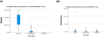
Serum HE4 levels among DKD, diabetic non-DKD, and healthy controls: serum HE4 levels differed significantly among the three study groups (p < 0.001). Patients with DKD exhibited markedly elevated HE4 levels compared with both diabetic patients without kidney disease and healthy controls. Post-hoc analysis confirmed significantly higher HE4 concentrations in the DKD group compared with the diabetes group (p < 0.001) and control group (p < 0.001). In addition, HE4 levels were significantly higher in diabetic patients compared with controls (p < 0.001). As illustrated in Figure 1, the DKD group demonstrated the highest median HE4 concentration with wide interquartile variability, whereas healthy controls showed consistently low HE4 levels. These findings demonstrate that serum HE4 levels differ significantly according to DKD status (Table 2, Figure 1).
Serum miR-615-3p expression according to DKD status: serum miR-615-3p expression differed significantly across the study groups (p = 0.002). The DKD group showed the highest expression levels, followed by the diabetes group and healthy controls. However, post-hoc analysis demonstrated that miR-615-3p expression did not differ significantly between the DKD and diabetes groups (p = 0.537). Although miR-615-3p expression was upregulated in DKD patients compared with controls, its standalone diagnostic performance appeared limited. Nevertheless, its combined assessment with HE4 may improve biomarker-based evaluation of DKD (Table 2, Figure 1).
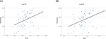
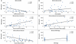
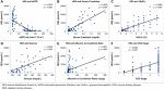
Correlation between HE4 and miR-615-fold regulation with other variables: in the DKD and diabetes groups, serum HE4 showed significant positive correlations with serum creatinine, albumin-to-creatinine ratio, CKD stage, HbA1c, and glucose levels, while demonstrating a significant inverse correlation with eGFR. These findings indicate a close association between HE4 and both renal dysfunction and glycemic control (Table 3, Figure 2, Figure 3, Figure 4). In contrast, miR-615-3p expression demonstrated limited significant correlations with clinical and biochemical parameters across the studied groups.
Main results: association of biomarkers with renal function parameters: to evaluate whether serum HE4 and miR-615-3p levels are associated with renal function parameters, receiver operating characteristic (ROC) curve analysis and correlation analyses were performed (Table 4).
Diagnostic performance of HE4 and miR-615-3p for DKD detection ROC analysis of individual and combined biomarkers: to address the study objective of evaluating the diagnostic utility of serum HE4 and miR-615-3p in diabetic kidney disease (DKD), receiver operating characteristic (ROC) curve analysis was performed to assess the discriminatory ability of each biomarker individually and in combination. Receiver operating characteristic analysis demonstrated high diagnostic accuracy of serum HE4 in distinguishing DKD among diabetic patients, with an area under the curve (AUC) of 0.969 (95% CI: 0.941–0.996, p < 0.001). At a cut-off value of 7,700 pmol/L, HE4 achieved 87.8% sensitivity and 94% specificity. In comparison, the albumin-to-creatinine ratio (ACR) demonstrated an AUC of 1.000 (95% CI: 1.000–1.000, p < 0.001), demonstrating the strongest diagnostic performance among the evaluated biomarkers in this cohort (Table 5, Figure 5). In contrast, miR-615-3p expression showed limited standalone diagnostic performance, with an AUC of 0.589 (95% CI: 0.476–0.702, p = 0.124), indicating poor discriminatory ability between DKD and non-DKD diabetic patients. Combined biomarker analysis demonstrated improved diagnostic performance. The combination of HE4 and miR-615-3p yielded an AUC of 0.972 (95% CI: 0.947–0.997, p < 0.001), with 87.8% sensitivity and 94% specificity, suggesting that they may serve as a complementary biomarker for DKD evaluation and monitoring. Additional ROC analysis was performed to evaluate the association between serum HE4 levels and advanced CKD stages (stages 3-5). HE4 demonstrated good discriminatory performance with an AUC of 0.969 (p < 0.001), while eGFR retained the highest diagnostic accuracy for CKD staging.
Independent predictors of DKD and clinical association analysis: logistic regression analysis was performed to determine whether HE4 and miR-615-3p were independently associated with DKD, as well as to investigate the clinical association between the studied biomarkers and CKD. Elevated log-transformed HE4 levels were significantly associated with the presence of CKD (OR = 145.046, 95% CI: 16.054-475.697, p < 0.001). Similarly, higher log-transformed miR-615-3p expression showed a significant association with CKD (OR= 11.868, 95% CI: 1.088-129.395, p= 0.042) (Table 6, Figure 5). These findings suggest that HE4 may serve as a promising complementary biomarker for DKD detection and monitoring, while miR-615-3p may provide additional diagnostic value when combined with HE4.
This study demonstrated significant differences in renal and metabolic profiles between patients with diabetic kidney disease (DKD), diabetic patients without kidney involvement, and healthy controls. Patients with DKD exhibited markedly elevated serum HE4 levels, together with worsening renal function parameters and albuminuria, supporting the potential role of HE4 as a biomarker reflecting renal injury and fibrosis in DKD. Human epididymis protein-4 is encoded by the whey acidic protein four-disulfide core domain 2 (WFDC2) gene and has been increasingly recognized as a marker of renal fibrosis and tubular injury in chronic kidney disease [22]. Experimental evidence suggests that HE4 contributes to extracellular matrix remodeling and fibrosis through inhibition of type I collagen degradation and promotion of inflammatory pathways [23,24]. In agreement with our findings, Song et al. [25] reported significantly elevated serum HE4 levels in CKD patients, with positive associations with disease severity and prognosis. Similarly, Zhang et al. [26] demonstrated significantly higher serum HE4 levels in DKD patients compared with diabetic patients without nephropathy and healthy controls. Comparable findings were also reported by Yuan et al. 2017 [27], who found significantly increased serum HE4 levels in CKD patients compared with controls. The present study also demonstrated significantly elevated serum miR-615-3p expression in DKD patients. However, unlike HE4, miR-615-3p showed considerable inter-individual variability and limited standalone diagnostic performance.
These findings suggest that miR-615-3p may be more closely related to pathogenic and regulatory mechanisms involved in fibrosis and inflammation rather than serving as an independent diagnostic marker. Previous studies have linked miR-615-3p dysregulation to renal fibrosis and diabetic complications. Wang et al. [8] demonstrated significantly increased urinary exosomal miR-615-3p expression in DKD patients and suggested its involvement in renal fibrotic processes [28]. Similarly, Bielska et al. [29] reported upregulation of serum miR-615-3p in diabetic patients with ischemic heart disease, supporting its potential role in diabetic complications. Experimental evidence further suggests that miR-615-3p may contribute to fibrosis progression through modulation of TGF-β signaling and epithelial-mesenchymal transition pathways [30]. Correlation analyses demonstrated significant associations between serum HE4 and markers of renal dysfunction and glycemic control, including serum creatinine, albumin-to-creatinine ratio, CKD stage, HbA1c, and glucose levels. These findings support the hypothesis that HE4 reflects both renal impairment and metabolic stress in diabetic patients. Similar associations between serum HE4 and declining renal function have been reported previously [26-27]. In contrast, miR-615-3p showed limited correlations with renal and metabolic parameters, suggesting that HE4 and miR-615-3p may represent distinct biological pathways in DKD pathogenesis.
ROC curve analysis demonstrated high diagnostic accuracy of serum HE4 for DKD detection, whereas miR-615-3p alone showed poor discriminatory ability. Zhang et al. [26] similarly demonstrated high diagnostic performance of serum HE4 for DKD detection with an AUC of 0.917. Yan et al. [31] also reported satisfactory diagnostic performance of HE4 in differentiating CKD patients and CKD stages. In contrast, Wang et al. [8] suggested that urinary exosomal miR-615-3p may have diagnostic value in DKD progression and renal fibrosis assessment. The discrepancy between their findings and the present study may be related to differences in specimen type, as urinary exosomal miRNAs may better reflect kidney-specific molecular changes than circulating serum miRNAs [32]. Combined biomarker analysis in the current study demonstrated improved diagnostic performance compared with miR-615-3p alone, supporting the potential value of multi-marker strategies in DKD evaluation. However, the albumin-to-creatinine ratio remained the strongest diagnostic marker in the current cohort, reaffirming its established role as the standard clinical marker for DKD detection. These findings are supported by a recent systematic review and meta-analysis by Jiang et al. [33], which concluded that individual microRNAs generally demonstrate limited standalone diagnostic performance, whereas integrated biomarker panels provide superior diagnostic accuracy for early DKD detection. Although the albumin-to-creatinine ratio (ACR) demonstrated the highest diagnostic performance in the present cohort, the objective of evaluating HE4 and miR-615-3p was not to replace established clinical markers. Rather, these biomarkers may provide complementary information regarding renal fibrosis and disease progression. Therefore, their potential clinical value may reside in risk stratification and mechanistic assessment rather than routine first-line diagnosis. Further longitudinal and cost-effectiveness studies are required before recommending their incorporation into routine clinical practice.
Logistic regression analysis further demonstrated significant associations between elevated HE4 and miR-615-3p levels and the presence of CKD. These findings are consistent with previous evidence linking HE4 to renal fibrosis and declining kidney function [26,28]. Recent studies have suggested that HE4 may contribute to extracellular matrix remodeling and renal fibrosis progression [28-34]. In addition, experimental studies have shown that miR-615 upregulation may promote renal interstitial fibrosis through increased fibronectin expression [8,35]. This study has several limitations. The relatively small sample size and single-center design may limit the generalizability of the findings. The study design precludes conclusions regarding causality or temporal changes in biomarker expression during DKD progression. The absence of histopathological assessment also limits direct evaluation of the relationship between biomarker expression and renal fibrosis severity. Furthermore, the limited diagnostic performance of serum miR-615-3p suggests that it may not be suitable as a standalone biomarker. A further limitation of this study is that eGFR was estimated using the MDRD equation rather than the Chronic Kidney Disease Epidemiology Collaboration (CKD-EPI) equation. Although MDRD has been widely used in clinical practice and research, CKD-EPI is currently recommended by contemporary guidelines because of its improved accuracy, particularly at higher levels of kidney function. Therefore, some degree of eGFR misclassification cannot be completely excluded. Future multicenter longitudinal studies with larger cohorts are warranted to validate these findings, clarify the mechanistic roles of HE4 and miR-615-3p in DKD progression, and explore their potential utility within integrated biomarker panels for early DKD detection and risk stratification.
Our findings highlight the importance of HE4 as a promising complementary biomarker for the diagnosis of DKD. Moreover, miR-615-3p could enhance the diagnostic accuracy for DKD detection when combined with HE4. Further large-scale studies are warranted to validate the clinical utility of these biomarkers for the detection of DKD.
What is known about this topic
- Diabetic kidney disease (DKD) is a major cause of chronic kidney disease and end-stage renal disease worldwide;
- Traditional renal biomarkers, including serum creatinine and albuminuria, may not adequately detect early renal injury or fibrosis in DKD;
- Biomarkers associated with renal fibrosis and inflammation, including Human Epididymis Protein 4 (HE4) and microRNAs, have been proposed as potential complementary tools for DKD detection and monitoring.
What this study adds
- Serum HE4 and serum miR-615-3p expression levels were significantly elevated in patients with DKD compared with diabetic patients without kidney disease and healthy controls;
- Serum HE4 demonstrated excellent diagnostic performance for DKD detection, while miR-615-3p showed limited standalone diagnostic value but may provide complementary value when combined with HE4;
- Elevated HE4 and miR-615-3p levels were significantly associated with chronic kidney disease among diabetic patients, supporting their potential role as non-invasive biomarkers for DKD detection and risk stratification.
The authors declare no competing interests.
Doaa Mamdouh Aly: conceptualization, methodology, data curation, formal analysis, investigation, writing original draft, review and editing. Asmaa Mohamed Fteah: conceptualization, methodology, formal analysis, investigation, data curation, validation, writing -review nd editing. Nihal M El-Assaly, Osama Mosbah: writing- review and editing. All authors have read and agreed to the final manuscript.
The authors would like to acknowledge the staff members of Theodor Bilharz Research Institute (TBRI) for their administrative support during the conduct of this study.
Table 1: demographic and laboratory data of the studied groups
Table 2: serum HE4 and miR-615 expression in the studied groups
Table 3: correlation of serum HE4 and eGFR with clinical/biochemical parameters in diabetic kidney disease, diabetes, and all patients
Table 4: correlation between eGFR and clinical parameters in the control group and all patients
Table 5: receiver operating characteristic curve analysis of HE4, serum miR-615 fold-change, microalbumin/creatinine ratio, eGFR, and their combinations for diabetic kidney disease and advanced chronic kidney disease prediction
Table 6: association of serum HE4 and miR-615-3p expression with chronic kidney disease risk among diabetic patients
Figure 1: comparison of serum human epididymis protein 4 and miR-615-3p expression levels among patients with diabetic kidney disease (DKD), diabetic patients without kidney disease, and healthy controls included in the study population; A) serum HE4 levels among the study groups; B) Serum miR-615-3p expression levels among the study groups; statistical significance was considered at p < 0.05
Figure 2: correlation analysis between serum human epididymis protein 4 levels and glycemic parameters among patients with diabetic kidney disease (DKD); A) correlation between serum HE4 levels and serum glucose levels among DKD patients; B) correlation between serum HE4 levels and HbA1c among DKD patients; pearson correlation analysis was performed. Statistical significance was considered at p < 0.05
Figure 3: correlation analysis between serum human epididymis protein 4 levels and renal function parameters among diabetic patients without diabetic kidney disease; A) correlation between serum HE4 levels and eGFR among diabetic patients; B) correlation between serum HE4 levels and serum urea among diabetic patients; C) correlation between serum HE4 levels and serum creatinine among diabetic patients; D) Correlation between serum HE4 levels and albumin-to-creatinine ratio among diabetic patients. E) correlation between serum HE4 levels and serum albumin among diabetic patients; F) correlation between serum HE4 levels and CKD stage among diabetic patients; pearson correlation analysis was performed. Statistical significance was considered at p < 0.05
Figure 4: corelation analysis between serum human epididymis protein 4 levels and clinical/biochemical parameters in the combined patient cohort (DKD and diabetes groups); A) Correlation between serum HE4 levels and eGFR; B) correlation between serum HE4 levels and serum creatinine; C) correlation between serum HE4 levels and HbA1c; D) correlation between serum HE4 levels and glucose levels; E) correlation between serum HE4 levels and albumin-to-creatinine ratio; F) correlation between serum HE4 levels and CKD stage. Pearson correlation analysis was performed. Statistical significance was considered at p < 0.05
Figure 5: receiver operating characteristic (ROC) curve analyses evaluating the diagnostic performance of serum HE4, serum miR-615-3p, and combined biomarker panels for diabetic kidney disease (DKD) and advanced chronic kidney disease (CKD); A) receiver operating characteristic analysis of serum HE4 as a biomarker for DKD detection; B) receiver operating characteristic analysis of serum miR-615-3p as a biomarker for DKD detection; C) receiver operating characteristic analysis of combined serum HE4 and serum miR-615-3p for DKD detection; D) receiver operating characteristic analysis comparing the combined HE4/miR-615-3p panel with albumin-to-creatinine ratio in DKD diagnosis; E) receiver operating characteristic analysis of serum HE4 for identifying advanced CKD stages (stages 3-5) against eGFR among all diabetic patients (DKD and diabetes groups)
- Rico-Fontalvo J, Aroca-Martínez G, Daza-Arnedo R, Cabrales J, Rodríguez-Yanez T, Cardona-Blanco M et al. Novel biomarkers of diabetic kidney disease. Biomolecules. 2023 Mar 31;13(4):633. 3. PubMed | Google Scholar
- Dianna JM, Edward JB. International Diabetes Federation Diabetes Atlas 10th edition. Accessed on September 14, 2025.
- Gheith O, Farouk N, Nampoory N, Halim MA, Al-Otaibi T. Diabetic kidney disease: worldwide difference of prevalence and risk factors. J Nephropharmacol. 2015 Oct 9;5(1):49-56. PubMed | Google Scholar
- Jung CY, Yoo TH. Pathophysiologic mechanisms and potential biomarkers in diabetic kidney disease. Diabetes Metab J. 2022 Mar;46(2):181-197. PubMed | Google Scholar
- Jha R, Lopez-Trevino S, Kankanamalage HR, Jha JC. Diabetes and renal complications: an overview on pathophysiology, biomarkers and therapeutic interventions. Biomedicines. 2024 May 15;12(5):1098. PubMed | Google Scholar
- Sakuma H, Hagiwara S, Kantharidis P, Gohda T, Suzuki Y. Potential targeting of renal fibrosis in diabetic kidney disease using microRNAs. Front Pharmacol. 2020 Nov 13:11:587689. PubMed | Google Scholar
- Mukhadi S, Hull R, Mbita Z, Dlamini Z. The role of microRNAs in kidney disease. Noncoding RNA. 2015 Nov 18;1(3):192-221. PubMed | Google Scholar
- Wang J, Tao Y, Zhao F, Liu T, Shen X, Zhou L. Expression of urinary exosomal miRNA-615-3p and miRNA-3147 in diabetic kidney disease and their association with inflammation and fibrosis. Ren Fail. 2023 Dec;45(1):2121929. PubMed | Google Scholar
- Yang B, Xie R, Wu SN, Gao CC, Yang XZ, Zhou JF. MicroRNA-615-5p targets insulin-like growth factor 2 and exerts tumor-suppressing functions in human esophageal squamous cell carcinoma. Oncol Rep. 2018 Jan;39(1):255-263. PubMed | Google Scholar
- International Diabetes Federation. International Diabetes Federation Diabetes Atlas 11th edition. Accessed on September 14, 2025.
- Levin A, Stevens PE, Bilous RW, Coresh J, De Francisco AL, De Jong PE et al. Kidney Disease: Improving Global Outcomes (KDIGO), CKD Work Group. KDIGO 2012 clinical practice guideline for the evaluation and management of chronic kidney disease. Kidney Int Suppl. 2013;3(1):1-150. Google Scholar
- American Diabetes Association Professional Practice Committee. 2 Diagnosis and Classification of Diabetes: Standards of Care in Diabetes-2024. Diabetes Care. 2024 Jan 1;47(Suppl 1):S20-S42. PubMed | Google Scholar
- American Diabetes Association. 2 Classification and Diagnosis of Diabetes: Standards of Medical Care in Diabetes-2021. Diabetes Care. 2021 Jan;44(Suppl 1):S15-S33. PubMed | Google Scholar
- Levey AS, Bosch JP, Lewis JB, Greene T, Rogers N, Roth D. A more accurate method to estimate glomerular filtration rate from serum creatinine: a new prediction equation. Ann Intern Med. 1999 Mar 16;130(6):461-70. PubMed | Google Scholar
- de Boer RA, Cao Q, Postmus D, Damman K, Voors AA, Jaarsma T et al. The WAP four-disulfide core domain protein HE4: a novel biomarker for heart failure. JACC Heart Fail. 2013 Apr;1(2):164-9. PubMed | Google Scholar
- Mestdagh P, Feys T, Bernard N, Guenther S, Chen C, Speleman F et al. High-throughput stem-loop RT-qPCR miRNA expression profiling using minute amounts of input RNA. Nucleic Acids Res. 2008 Dec;36(21):e143. PubMed | Google Scholar
- Livak KJ, Schmittgen TD. Analysis of relative gene expression data using real-time quantitative PCR and the 2(-Delta Delta C(T)) method. Methods. 2001 Dec;25(4):402-8. PubMed | Google Scholar
- Chan YH. Biostatistics 102: quantitative data-parametric and non-parametric tests. Singapore Med J. 2003 Aug;44(8):391-6. PubMed | Google Scholar
- Chan YH. Biostatistics 103: qualitative data-tests of independence. Singapore Med J. 2003 Oct;44(10):498-503. PubMed | Google Scholar
- Chan YH. Biostatistics 104: correlational analysis. Singapore Med J. 2003;44(12):614-619. PubMed | Google Scholar
- Chan YH. Biostatistics 202: logistic regression analysis. Singapore Med J. 2004 Apr;45(4):149-53. PubMed | Google Scholar
- Smith HO, Berwick M, Verschraegen CF, Wiggins C, Lansing L, Muller CY et al. Incidence and survival rates for female malignant germ cell tumors. Obstet Gynecol. 2006 May;107(5):1075-85. PubMed | Google Scholar
- Wang J, Zhao H, Xu F, Zhang P, Zheng Y, Jia N. Human epididymis protein 4 (HE4) protects against cystic pulmonary fibrosis-associated inflammation through inhibition of NF-?B and MAPK signaling. Genes Genomics. 2019 Sep;41(9):1045-1053. PubMed | Google Scholar
- An C, Jiao B, Du H, Tran M, Song B, Wang P et al. Jumonji domain-containing protein-3 (JMJD3) promotes myeloid fibroblast activation and macrophage polarization in kidney fibrosis. Br J Pharmacol. 2023 Sep;180(17):2250-2265. PubMed | Google Scholar
- Song J, Chen L, Yuan Z, Gong X. Elevation of serum human epididymis protein 4 (HE4) and N-terminal pro-B-type natriuretic peptide (NT-proBNP) as predicting factors for the occurrence of acute kidney injury on chronic kidney disease: a single-center retrospective self-control study. Front Pharmacol. 2023 Sep 11;14:1269311. PubMed | Google Scholar
- Zhang M, Zhao B, Xie J, Liang Y, Yang Z. Serum human epididymis protein 4 is associated with renal function and diabetic kidney disease in patients with type 2 diabetes mellitus. Biomed Res Int. 2019 Nov 11;2019:4831459. PubMed | Google Scholar
- Yuan T, Li Y. Human epididymis protein 4 as a potential biomarker of chronic kidney disease in female patients with normal ovarian function. Lab Med. 2017 Aug 1;48(3):238-243. PubMed | Google Scholar
- Wan J, Wang Y, Cai G, Liang J, Yue C, Wang F et al. Elevated serum concentrations of HE4 as a novel biomarker of disease severity and renal fibrosis in kidney disease. Oncotarget. 2016 Oct 18;7(42):67748-67759. PubMed | Google Scholar
- Bielska A, Niemira M, Bauer W, Sidorkiewicz I, Szałkowska A, Skwarska A et al. Serum miRNA profile in diabetic patients with ischemic heart disease as a promising non-invasive biomarker. Front Endocrinol (Lausanne). 2022 May 18;13:888948. PubMed | Google Scholar
- Lei B, Wang D, Zhang M, Deng Y, Jiang H, Li Y. miR-615-3p promotes the epithelial-mesenchymal transition and metastasis of breast cancer by targeting PICK1/TGFBR1 axis. J Exp Clin Cancer Res. 2020 Apr 26;39(1):71. PubMed | Google Scholar
- Yan S, Lin Y, Tian X. Significantly elevated serum human epididymis protein-4 in chronic kidney disease patients without ovarian cancer: a large-scale retrospective study. J Clin Lab Anal. 2023 Apr;37(4):e24847. PubMed | Google Scholar
- Tang Y, Hu Z, Liu Z, Peng S, Liu T, Xiao Y et al. HE4 serum levels are associated with poor prognosis in patients with acute heart failure combined with chronic kidney disease. Int J Gen Med. 2024 Apr 2;17:1273-1280. PubMed | Google Scholar
- Jiang L, Li L, Zhang K, Zheng L, Zhang X, Hou Y et al. A systematic review and meta-analysis of microRNAs in the diagnosis of early diabetic kidney disease. Front Endocrinol (Lausanne). 2025 Apr 22;16:1432652. PubMed | Google Scholar
- LeBleu VS, Teng Y, O'Connell JT, Charytan D, Müller GA, Müller CA et al. Identification of human epididymis protein-4 as a fibroblast-derived mediator of fibrosis. Nat Med. 2013 Feb;19(2):227-31. PubMed | Google Scholar
- Zhu Y, Wang Z, Liang Z, Xu S, Teng Y, Li X et al. LncRNA4474 inhibits renal fibrosis by regulating hepatocyte nuclear factor-1β through miR-615 modulation. Cell Cycle. 2022 Jun;21(12):1233-1248. PubMed | Google Scholar
Search
This article authors
On Pubmed
On Google Scholar
Citation [Download]
Navigate this article
Similar articles in
Key words
Tables and figures


Article metrics
Recently from the PAMJ






Authors´ services
