
Intraoperative diagnosis and management of gastric ischemia: a rare case report and literature review
Ahmed Hadj Taieb, Sami Fendri, Majdi Kchaw, Youssef Mejdoub, Haitham Ben Rejab, Salah Boujelben
Corresponding author: Department of General Surgery, Habib Bourguiba University Hospital, Sfax, 3000, Tunisia 
Received: 18 Nov 2025 - Accepted: 04 Aug 2026 - Published: 02 Sep 2026
Domain: Radiology,Gastroenterology,General surgery
Keywords: Gastric ischemia, mesenteric ischemia, atypical gastrectomy, gastric wall necrosis, case report
Funding: This work received no specific grant from any funding agency in the public, commercial, or non-profit sectors.
©Ahmed Hadj Taieb et al. Pan African Medical Journal (ISSN: 1937-8688). This is an Open Access article distributed under the terms of the Creative Commons Attribution International 4.0 License (https://creativecommons.org/licenses/by/4.0/), which permits unrestricted use, distribution, and reproduction in any medium, provided the original work is properly cited.
Cite this article: Ahmed Hadj Taieb et al. Intraoperative diagnosis and management of gastric ischemia: a rare case report and literature review. Pan African Medical Journal. 2026;55:1. [doi: 10.11604/pamj.2026.55.1.50271]
Available online at: https://www.panafrican-med-journal.com//content/article/55/1/full
 PDF in process
 Case report 

Intraoperative diagnosis and management of gastric ischemia: a rare case report and literature review
Intraoperative diagnosis and management of gastric ischemia: a rare case report and literature review
Ahmed Hadj Taieb1,&,  Sami Fendri1, Majdi Kchaw1, Youssef Mejdoub1, Haitham Ben Rejab1, Salah Boujelben1
&Corresponding author
Gastric ischemia (GI) is an exceptionally rare condition due to the stomach's rich collateral circulation, making early diagnosis challenging. We report the case of a 64-year-old man with multiple cardiovascular comorbidities who presented with acute abdominal pain and diarrhea. Despite initial medical treatment, his condition worsened, leading to emergency exploratory laparotomy. Intraoperatively, ischemic "map-like" lesions of the posterior gastric wall were discovered, prompting an atypical gastrectomy given the patient's hemodynamic instability and the impossibility of revascularization. Postoperative evolution was marked by cardiac decompensation followed by cardiopulmonary arrest. This case highlights the importance of maintaining a high index of suspicion for gastric ischemia in high-risk patients and illustrates the diagnostic challenges when ischemic changes are only identified intraoperatively. Reporting such rare presentations may help improve understanding, early recognition, and management of this life-threatening condition.

Mesenteric ischemia can result from either arterial or venous insufficiency or may occur non-occlusively [1]. Several factors predispose individuals to this condition, including cardiovascular diseases, smoking, atrial fibrillation, hypercoagulability, and vasculitis [2]. On the other hand, gastric ischemia (GI) is relatively rare due to the stomach's extensive collateral blood supply. The infrequency of GI is underscored by a comprehensive retrospective study on ischemic gastritis conducted from January 2000 to May 2016, which identified only 17 cases of isolated gastric ischemia at a single center [3]. This rarity contributes to the absence of a standardized treatment protocol. Here, we present a case of gastric ischemia identified during intraoperative examination at our academic institution.

Patient information: a 64-year-old male with a medical history of diabetes mellitus, end-stage renal disease on hemodialysis, and coronary artery disease with coronary stenting was brought by ambulance to the emergency department for abdominal pain associated with diarrhea evolving over four days. There was no previous history of abdominal surgery or known chronic gastrointestinal disease reported.
Timeline of the current episode: four days before admission, the patient developed acute abdominal pain and seven episodes of diarrhea. He consulted the emergency department, where initial clinical assessment and laboratory work-up were performed. Despite initiation of medical treatment, his abdominal pain and general condition worsened within 24 hours, leading to the indication for emergency exploratory laparotomy.
Clinical findings: on admission, the patient was afebrile, hemodynamically and respiratory stable. Abdominal examination revealed a soft, depressible abdomen without guarding or peritoneal signs. Digital rectal examination showed normochromic, liquid stools. There were no signs of overt gastrointestinal bleeding.
Diagnostic assessment: laboratory tests demonstrated a marked inflammatory response with a C-reactive protein (CRP) level of 435 mg/L and a white blood cell count of 31,000/mm3. Contrast-enhanced abdominopelvic computed tomography showed extensive atheromatous and calcified infiltration of the abdominal aorta and its branches, including a calcified plaque at the origin of the celiac trunk with preserved patency of the hepatic artery. Massive calcification of the splenic artery was noted, which was occluded 4 cm from its origin, associated with multiple non-enhancing hypodense areas in the mid-splenic and lower polar regions, consistent with splenic infarctions. The superior and inferior mesenteric arteries and their branches also showed diffuse atheromatous and calcified involvement, with some segments appearing severely narrowed. A mild circumferential wall thickening of the sigmoid colon was observed, suggestive of an infectious process. No specific gastric wall abnormality was initially identified.
Diagnosis: in view of the persistent abdominal pain, inflammatory syndrome, extensive splanchnic vascular disease on imaging, and subsequent intraoperative findings, the final diagnosis was acute gastric ischemia localized to the posterior wall of the stomach in a patient with severe multivessel atherosclerotic disease.
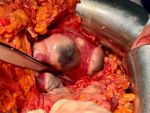
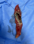
Therapeutic interventions: given the worsening abdominal pain and the onset of diffuse abdominal tenderness despite antibiotic therapy, the patient was taken to the operating room 24 hours after admission for exploratory laparotomy. Intraoperatively, the small bowel and the entire colonic frame appeared normal. Fibrinous membranes were observed over the gastrocolic ligament. Dissection of the gastrocolic ligament and entry into the lesser sac revealed ischemic lesions on the posterior wall of the stomach, with a "map-like" appearance in a pre-perforative stage (Figure 1). The pancreas was otherwise normal. Due to the patient's hemodynamic instability and the need for high doses of catecholamines, revascularization was not feasible. An atypical gastrectomy of the affected posterior gastric wall was therefore performed using a surgical stapler (Figure 2).
Follow-up and outcomes: the patient was transferred to the intensive care unit postoperatively. Early postoperative evolution was unfavorable, marked by cardiac decompensation followed by cardiopulmonary arrest, leading to death despite resuscitation efforts.
Patient perspective: according to the patient's next of kin, the family understood the severity of his condition and appreciated the efforts of the medical and surgical teams. They consented to publication of this case in the hope that it may help improve the recognition and management of similar situations in other patients.
Informed consent: written informed consent for publication of this case report and accompanying images was obtained from the patient's next of kin.
Gastric ischemia is an exceptionally rare condition with limited documentation in medical literature, primarily found in a small number of case reports and series. Its rarity is largely due to the stomach's rich collateral circulation, which includes the celiac trunk, superior mesenteric artery, and other collateral vessels. The stomach's strong submucosal vascular plexus offers greater resistance to ischemia than the intestines. Despite this, GI can still occur as a result of both localized and widespread vascular insufficiency, which may be due to various causes. These causes can be classified into systemic hypoperfusion (such as shock or sepsis) or hypoperfusion of splanchnic vessels due to conditions like gastric volvulus, acute gastric dilatation, stenosis, thrombosis, embolism, vasculitis or vasoconstriction [1,4]. Rarely, GI may also stem from endoscopic procedures (such as submucosal dissection or injection sclerotherapy), postoperative situations (e.g., distal pancreatectomy with celiac axis resection, subtotal gastrectomy, highly selective vagotomy, splenectomy, gastric restrictive surgery), or even substance abuse like [1,4]. Several risk factors, including older age, smoking, atherosclerosis, diabetes, and hypertension, can reduce blood flow to the stomach and splanchnic vessels, heightening the risk of ischemia [5].
Clinically, GI presents with different symptoms based on whether the condition is acute or chronic. In the acute phase, patients may experience nausea, vomiting, upper gastrointestinal bleeding, abdominal distention, and symptoms indicative of underlying conditions such as shock or hypoperfusion. Chronic GI can manifest as ischemic gastroparesis, with patients showing abdominal pain, nausea, vomiting, gastrointestinal bleeding (either overt or hidden), anemia, diarrhea, and weight loss [1,4]. Imaging techniques, especially CT scans, play a crucial role in diagnosing GI, with common signs including gastric wall pneumatosis and portal venous gas [6]. However, these findings are not specific to GI since bacterial infections or increased intragastric pressures can present similarly [3]. While CT can sometimes identify underlying causes, it does not rule out GI, as endoscopy is more sensitive in detecting early ischemic changes. Endoscopic findings include mucosal congestion, a red or purplish hue, large ulcerations (with or without bleeding), and necrosis with black/gray eschar, often located where arterial branches connect the lesser and greater curvatures of the stomach [3].
In cases with vascular obstruction, angiographic treatments like angioplasty or stenting may be used to restore blood flow [7]. Treatment strategies depend on the severity of ischemia. Acute cases often require acid suppression and gastric decompression [1,3] , while broad-spectrum antibiotics are recommended for cases showing pneumatosis or gas in the portal system [1,3,8]. Conservative treatment can have a 24% mortality rate within six months, although recurrence of bleeding is rare [9]. When complications like perforation, sepsis, or persistent bleeding arise, surgical intervention, including gastrectomy, may be necessary [10]. In our case, GI was diagnosed during surgery, and due to the patient's hemodynamic instability, revascularization was not an option, leading to the decision for an atypical gastrectomy.
Gastric ischemia is an extremely rare condition, primarily due to the stomach's robust collateral blood supply, which makes it naturally resistant to ischemic injury. Its diagnosis is challenging and requires a combination of imaging and endoscopic evaluation. Management depends on the severity and underlying etiology, with no standardized protocols available due to its rarity. Despite advancements in care, the prognosis remains poor, particularly in cases complicated by hemodynamic instability or significant comorbidities. This highlights the need for early recognition, prompt intervention, and further research to develop evidence-based management strategies for this uncommon but serious condition.
The authors declare no competing interests.
Ahmed Hadj Taieb: conceptualization, patient management, and drafting. Sami Fendri: surgical management and resources. Majdi Kchaw: literature review, writing -revision and editing. Youssef Mejdoub: data analysis and methodology. Haitham Ben Rejab: revision and editing. Salah Boujelben: supervision, validation, and final approval. All the authors have read and approved the final version of this manuscript.
Figure 1: ischemic lesions on the posterior wall of the stomach
Figure 2: gastrectomy specimen
- Tang S, Daram SR, Wu R, Bhaijee F. Pathogenesis, diagnosis, and management of gastric ischemia. Clin Gastroenterol Hepatol. 2014;12(2):246-252. PubMed | Google Scholar
- Björck M, Koelemay M, Acosta S, Goncalves FB, Kölbel T, Kolkman JJ et al. Editor's choice-management of the diseases of mesenteric arteries and veins: clinical practice guidelines of the European Society of Vascular Surgery (ESVS). Eur J Vasc Endovasc Surg. 2017;53(4):460-510. PubMed | Google Scholar
- Sharma A, Mukewar S, Chari ST, Wong Kee Song LM. Clinical Features and Outcomes of Gastric Ischemia. Dig Dis Sci. 2017;62(12):3550-3556. PubMed | Google Scholar
- Herman J, Chavalitdhamrong D, Jensen D, Cortina G, Manuyakorn A, Jutabha R. The significance of gastric and duodenal histological ischemia reported on endoscopic biopsy. Endoscopy. 2011;43(04):365-368. PubMed | Google Scholar
- Casey KM, Quigley TM, Kozarek RA, Raker EJ. Lethal nature of ischemic gastropathy. Am J Surg. 1993;165(5):646-649. PubMed | Google Scholar
- Binmoeller KF, Benner KG. Emphysematous gastritis secondary to gastric infarction. Am J Gastroenterol Springer Nat. 1992;87(4). PubMed | Google Scholar
- Huang G, Jin Y. Total gastric necrosis: A case report and literature review. Niger J Clin Pract. 2017;20(5):645-646. PubMed | Google Scholar
- Acar T, Çakir V, Acar N, Atahan K, Haciyanli M. Chronic visceral ischemia: an unusual cause of abdominal pain. Turk J Surg. 2018;34(2):158. PubMed | Google Scholar
- Silva JC, Rodrigues A, Ponte A, Silva AP, Carvalho J. A case of gastric ischemia: management and prognosis. GE Port J Gastroenterol. 2020 Jan;27(1):53-55. PubMed | Google Scholar
- Dueñas CS, González AE, Lacunza AG. Gastric ischemia due to critical stenosis of the celiac trunk. IAn Sist Sanit Navar. 2018 Apr 30;41(1):123-127. PubMed | Google Scholar
Search
This article authors
On Pubmed
On Google Scholar
Citation [Download]
Navigate this article
Similar articles in
Key words
Tables and figures
Article metrics
Recently from the PAMJ




Authors´ services
