
Bedside ultrasonographic evaluation of pulmonary and diaphragmatic function following cardiac surgery and it's association with early respiratory outcomes: a prospective observational study
Bharti Sidhu, Banashree Mandal, Goverdhan Dutt Puri, Virendra Kumar Arya, Ashwini Reddy
Corresponding author: Banashree Mandal, Department of Anaesthesia and Intensive Care, Post Graduate Institute of Medical Education and Research, Chandigarh, India 
Received: 12 Oct 2025 - Accepted: 03 Jul 2026 - Published: 22 Jul 2026
Domain: Intensive care medicine,Cardiovascular surgery,Surgical Sciences
Keywords: Lung ultrasound, B-lines, diaphragm ultrasound, cardiopulmonary bypass, mechanical ventilation, weaning
Funding: This work received no specific grant from any funding agency in the public, commercial, or non-profit sectors.
©Bharti Sidhu et al. Pan African Medical Journal (ISSN: 1937-8688). This is an Open Access article distributed under the terms of the Creative Commons Attribution International 4.0 License (https://creativecommons.org/licenses/by/4.0/), which permits unrestricted use, distribution, and reproduction in any medium, provided the original work is properly cited.
Cite this article: Bharti Sidhu et al. Bedside ultrasonographic evaluation of pulmonary and diaphragmatic function following cardiac surgery and it's association with early respiratory outcomes: a prospective observational study. Pan African Medical Journal. 2026;54:93. [doi: 10.11604/pamj.2026.54.93.49700]
Available online at: https://www.panafrican-med-journal.com//content/article/54/93/full
 PDF in process
 Research 

Bedside ultrasonographic evaluation of pulmonary and diaphragmatic function following cardiac surgery and it's association with early respiratory outcomes: a prospective observational study
Bedside ultrasonographic evaluation of pulmonary and diaphragmatic function following cardiac surgery and it's association with early respiratory outcomes: a prospective observational study
 Bharti Sidhu1, Banashree Mandal2,&, Goverdhan Dutt Puri3, Virendra Kumar Arya4, Ashwini Reddy2
&Corresponding author
Introduction: postoperative pulmonary complications are common after cardiac surgery, influenced by cardiopulmonary bypass, fluid shifts, and diaphragmatic impairment. Bedside ultrasonography can assess extravascular lung water and diaphragm function, and may aid in ventilatory support decisions. This study evaluated lung and diaphragm ultrasonographic changes following cardiac surgery and their association with weaning outcomes.
Methods: in this prospective observational study, adult patients aged ≥18 years undergoing elective cardiac surgery with cardiopulmonary bypass were assessed using lung and diaphragm ultrasound preoperatively, during minimal pressure support ventilation (PSV) with 5 cm H2O of PEEP and 5 cm H2O of pressure support, and four hours post-extubation. The primary outcome was weaning failure, defined as the need for non-invasive ventilation (NIV) or reintubation within 48 hours. Parameters included B lines, lung ultrasound scores (LUS), diaphragmatic excursion, and thickness.
Results:among 73 patients, 57 (78 %) had weaning success, and 13 ( 17.80% ) had weaning failure (required NIV). The weaning failure group was older (56.69±13.70 vs. 45.65±14.84 years; P=0.014), had higher body mass index (BMI) (27.11±5.37 vs. 21.92±4.06 kg/m2; P=0.002), more comorbidities (76.9% vs. 41.7%; P=0.02), and more coronary artery disease (76.9% vs. 33.3%; P=0.006). Ultrasound showed increased B-lines and LUS and reduced diaphragmatic excursion postoperatively (P< 0.05) in both groups, but no significant differences between groups. B-lines correlated with prolonged intensive care unit (ICU) stays.
Conclusion: ultrasound detected postoperative lung and diaphragm changes, but did not independently predict weaning failure. Clinical factors such as age, body mass index (BMI), comorbidities, postoperative cardiac function, and Pa/FiO2 ratio were stronger predictors. Ultrasound should complement clinical evaluation in guiding weaning decisions.

Postoperative respiratory complications remain a significant concern in cardiac surgery patients, with about 2.6% to 22.7% of patients requiring prolonged mechanical ventilation [1]. Extubation failure is defined as the inability to sustain spontaneous breathing after removal of the endotracheal tube and the need for reintubation within 24-72 hours [2,3]. Incidence of extubation failure in cardiac surgical patients has been found to be 6.6% [4]. Postoperative respiratory complications are largely influenced by cardiopulmonary bypass (CPB)-induced inflammation, fluid shifts, surgical trauma, the effect of anaesthesia, and diaphragmatic dysfunction. Fluid shifts and systemic inflammatory response cause fluid to accumulate in alveolar and interstitial spaces. The increase in extravascular lung water impairs gas exchange and may lead to prolonged mechanical ventilation. Difficult weaning from mechanical ventilation and weaning failure have also been seen in patients with diaphragmatic dysfunction. Traditionally, clinical indices have been relied upon to guide weaning from mechanical ventilation. Bedside ultrasonography has emerged as a non-invasive method to directly assess lung water and diaphragmatic function; however, the predictive capability of bedside ultrasonography in cardiac surgical settings remains uncertain. B Lines are reverberation artifacts seen on a lung ultrasound scan.
Sonographic B lines are related to radiographic Kerley B-Lines and lung water score on chest X-ray, to extravascular lung water (EVLW) measured invasively by the thermodilution method [5,6]. Based on the presence and severity of B lines, the lung ultrasound score (LUS) is calculated, which can be a valuable tool in cardiac surgery patients for assessing lung aeration and hence aid in the prediction of postoperative respiratory complications [6]. Diaphragmatic dysfunction is commonly seen in post-cardiac surgery patients (incidence 1%-60%) secondary to surgical manipulation, traction, traumatic injury, and due to the use of topical cooling, which can be assessed with bedside ultrasound [7]. Hence, we conducted this study with the primary objective of finding the correlation of extravascular lung water (B lines) and diaphragmatic function (excursion, thickness) using ultrasonography with the weaning failure in adult post-cardiac surgery patients. Weaning failure was defined as the need for non-invasive ventilation (NIV) or reintubation within 48 hours of extubation during primary stay in the intensive care unit. The secondary objectives included the correlation of weaning failure predicted by ultrasonography with the more commonly used weaning index, that is, the PaO2/FiO2 ratio, duration of non-invasive ventilation during stay in the intensive care unit, and duration of intensive care unit stay. This study also aimed to clarify the clinical utility of ultrasonographic evaluations by examining their association with early postoperative respiratory outcomes following cardiac surgery.

Study design: this prospective observational study was conducted to evaluate lung and diaphragm ultrasonographic changes following cardiac surgery and their association with weaning outcomes.
Study setting and population: this study was conducted from January 2016 to June 2017 at a tertiary care center in North India. The study population included adult patients aged ≥ 18 years undergoing elective cardiac surgery with cardiopulmonary bypass. Exclusion criteria included emergency surgery, inadequate ultrasound windows, dressing at the scanning site, or refusal to participate.
All the patients received standardized general anesthesia with graded doses of intravenous fentanyl (2 mcg/kg), propofol titrated to loss of verbal response (1-2 mg/kg), and vecuronium (0.1 mg/kg) to facilitate endotracheal intubation. The cardiopulmonary bypass management included the use of normothermia, conventional ultrafiltration with a target hemoglobin of 8 gm/dl, and termination of cardiopulmonary bypass was done using appropriate doses of inotropes as deemed necessary by the attending anesthesiologist. After surgery, patients were shifted to the ICU and were mechanically ventilated with a tidal volume of 6-8 ml/kg, positive end expiratory pressure (PEEP) 5 mmHg, and an inspiratory to expiratory ratio of 1:2 (SIMV/PRVC mode, Servo-i ventilator; Maquet, Getinge AB, Getinge, Sweden). Patients were weaned from mechanical ventilation at the discretion of the attending anesthesiologist. During weaning, all patients were brought to pressure support ventilation (PSV) with 5 cm H2O of PEEP and 5 cm H2O of pressure support. The patients were evaluated for extubation readiness by the attending intensivist, who was blinded to the ultrasound results based on cardiovascular stability, minimal chest drain output, absence of neurological deficit, PaO2/FiO2 ratio (PF ratio) ≥200, and rapid shallow breathing index (RSBI) ≤150.
Data collection: ultrasound examinations of the lungs and diaphragm were conducted at three time points: preoperatively, during minimal PSV (with 5 cm H2O of PEEP and 5 cm H2O of pressure support), and four hours post-extubation.
Lung ultrasonography: lung ultrasound was performed in the supine position using a 3.5-5 MHz convex probe (Sonosite Fuzifilm M-Turbo). The thorax was divided into 12 zones for exploration, using anatomical landmarks for delimitation. On each side, the zones were bounded by the parasternal line, the anterior axillary line, the posterior axillary line, and a paravertebral zone. Vertically, the thorax was divided into upper and lower regions by a horizontal line drawn at the level of the nipples, perpendicular to the aforementioned lines. The lung ultrasound scan was performed, placing the focus at the level of the pleural line (2-4 cm) and setting the depth to 8 to 10 cm. A scan was performed in each of the 12 zones in the longitudinal plane, and the pattern of the least aeration present in each zone was assessed. In case of requiring a better ultrasonic window and/or better evaluation of the area, the transducer was placed in the transverse plane. A semi-quantitative lung ultrasound score ranging from 0 to 3 was calculated based on the number of B lines present: score 0: ≤ 5 B-lines, 1: 6-15 B-lines, 2: 16-20 B-lines, and 3: > 30 B-lines [6].
Diaphragm ultrasonography: diaphragmatic excursion was assessed using the 2-5 MHz curvilinear probe (Sonosite Fuzifilm M-Turbo) with the patient in the supine position. The right hemidiaphragm was evaluated with a probe placed between the midclavicular and axillary line, in the right subcostal area, and directed medially, cranially, and dorsally. The diaphragmatic movements were measured using M-mode [8]. Measurements were performed during quiet breathing. The left hemidiaphragm was studied with a probe placed over the left subcostal line or low intercostal position between the anterior and mid axillary lines. The diaphragm was seen as a single echogenic line, moving towards the probe during inspiration and away from the probe during expiration. Diaphragmatic excursion was defined as the difference between the highest point and the lowest point (amplitude). The diaphragm thickness was evaluated with a linear 6-13 MHz probe (Sonosite Fuzifilm M-Turbo) in the B-mode [9]. The diaphragm was identified as a three-layered structure with two parallel echogenic lines, comprising two hyperechoic outer layers (the peritoneum and pleura) surrounding a hypoechoic inner muscle layer. The thickness of each hemidiaphragm was determined by directly measuring the distance between the center of the pleural membrane and the center of the peritoneal membrane on the frozen B-mode images. The measurements were collected at the end of maximal inspiration and maximal expiration. Average measurements from at least three separate breathing cycles were taken.
Additional parameters and collection of outcomes: transthoracic echocardiography was performed at the same three time points to measure left ventricular ejection fraction (LVEF). Arterial blood gas analysis was used to calculate the PF (PaO2/FiO2) ratio taken at minimal PSV postoperatively. The requirement for non-invasive ventilation (NIV) or reintubation within 48 hours of extubation was recorded. Patients requiring either of these were considered to have experienced weaning failure.
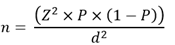
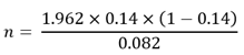
Sample size: the sample size was calculated using the Cochran formula, based on a previous study done by Soummer et al. (2012), [10] which reported a 14% incidence of extubation failure. Assuming a 95% confidence interval (Z=1.96) and a margin of error (d=0.08), the required sample size was calculated as:
Data analysis: all statistical analyses were performed using statistical package for the social sciences (SPSS) (version 22.0; SPSS Inc., Chicago, IL, USA). Normality of data was checked using the Shapiro-Wilk test. The variables on a continuous scale were described as mean ± standard deviation (SD) or median with interquartile range. The parametric data were compared with the Student t-test, and the non-parametric data were analyzed using the Mann-Whitney U test. All statistical tests were two-sided, and a P value of < 0.05 was considered significant.
Ethical consideration: this study was approved by the Institute Ethics Committee of the Post Graduate Institute of Medical Research in Chandigarh, India (IEC No: INT/IEC/2017/788). All adults ≥18 years of age were asked to give informed consent and sign a consent form before participating in the study. For participants who were unable to read, we read and explained the consent form to them. Confidentiality was ensured, and the electronic data has been stored in a password-protected computer.
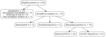
Socio-demographic analysis: of the 90 patients initially enrolled, 17 were excluded based on predefined criteria. The final analysis included 73 patients with a mean age of 47±15 years (Figure 1). Among these, two patients required reintubation, and one underwent tracheostomy. These three cases were excluded from the weaning failure analysis, as reintubation occurred due to non-respiratory causes, including hemodynamic instability and neurological impairment. Notably, their ultrasound parameters, including B-lines, lung ultrasound scores (LUS), diaphragmatic excursion, and diaphragmatic thickness, did not differ significantly from those of other patients. Demographic and clinical characteristics of the patients who failed weaning versus those who succeeded in the weaning trial are given in Table 1. Patients in the weaning failure group were significantly older (56.69±13.70 vs. 45.65±14.84 years; P=0.014) and had a higher body mass index (BMI) (27.11±5.37 vs. 21.92±4.06 kg/2P=0.002) as compared to the weaning success group. Comorbidities were more frequent in the failure group (76.9% vs. 41.7%; P=0.02), and coronary artery disease (CAD) was the predominant diagnosis in the weaning failure group (76.9% vs. 33.3%; P=0.006).
Assessment of primary outcome
Lung and diaphragm ultrasound parameters between groups: lung ultrasound findings, including B-line counts and lung ultrasound scores (LUS), did not significantly differ between the weaning success and weaning failure groups at three time points: baseline, during minimum pressure support ventilation (PSV), and during post-extubation. The B-line counts post-extubation were 21.88 ± 16.79 in the weaning success group versus 20.08 ±18.12 in the failure group (P = 0.376), and LUS values post-extubation were 1.67±0.95 vs. 1.46±1.05 (P = 0.773), Table 2. Diaphragmatic excursion measured on the right side was lower in the weaning failure group across all time points, with a near-significant difference at baseline (1.48±0.33 vs. 1.56±0.52cm; P=0.060) and post-extubation (1.04±0.52vs. 1.11±0.41cm; P=0.070). Left diaphragmatic excursion did not show consistent directional changes. Diaphragmatic thickness and diaphragmatic thickening fraction (DTF) parameters were comparable between groups ( Table 2). Transthoracic echocardiography revealed that patients had significantly lower left ventricular ejection fraction (LVEF) in the weaning failure group at both baseline (45.00±7.00 vs. 47.65±8.67%; P=0.029) and during PSV (43.00±7.81 vs. 48.15±8.11%; P=0.040), although post-extubation values were equivalent (P=0.883). A statistically significant difference in PaO2/FiO2 ratios was observed in the post-extubation period between patients who failed weaning and those who succeeded (203.61±60.79 vs. 247.84±54.95; P = 0.007), suggesting compromised gas exchange in the weaning failure group patients (Table 2).
Assessment of secondary outcomes
ICU stay and mechanical ventilation duration: a positive correlation was found between ICU stay duration and both cardiopulmonary bypass (CPB) time (correlation coefficient r=0.346, P=0.003) and aortic cross-clamp (AXC) time (r=0.258, P=0.03). This indicates that with an increase in CPB and AXC time, ICU stay duration increases. However, neither CPB nor AXC time showed a significant correlation with the duration of mechanical ventilation (P > 0.05). This suggests that while increased CPB and AXC durations are associated with longer ICU stays, they do not necessarily prolong ventilatory support.
Temporal trends in lung and diaphragmatic parameters: serial evaluations among three time points (baseline, minimum pressure support ventilation, and 4 hours post-extubation) revealed statistically significant changes in lung aeration and diaphragmatic function in all the patients (Table 3). All patients demonstrated a statistically significant increase in B-lines from baseline (11.34±17.66) to PSV (22.68±20.62) and remained elevated post-extubation (21.56±16.90), with P=0.01 for both transitions. LUS calculated from B-Line count also rose significantly from baseline to PSV (P=0.01) and to post-extubation (P=0.02). Right diaphragmatic excursion decreased from baseline (1.53±.0.49 cm) to PSV (1.25±0.46) and further to post-extubation (1.09±0.42), with significant drops observed in both comparisons (P=0.01 each). The left diaphragmatic excursion showed a decrease at PSV (0.96±0.32) compared to baseline (1.40±47), followed by partial recovery in the post-extubation period (1.37±10.49). These changes were statistically significant when compared to baseline.
Inspiratory and expiratory thickness of the diaphragm on both sides decreased significantly from baseline to both the PSV and post-extubation periods. Right inspiratory thickness fell from 0.55±.0.23 at baseline to 0.46±0.17 during PSV (P=0.01), returning to baseline values post-extubation. Expiratory thickness showed a persistent reduction throughout (P<0.01). Similar patterns were observed on the left side, Table 3. Among intraoperative variables, cardiopulmonary bypass (CPB) duration was significantly longer in the weaning failure group (201.46±141.50 vs. 139.03±66.21 minutes; P=0.049). There was no significant difference in aortic cross-clamp time and lowest core temperature between the two groups. Non-invasive ventilation was used only in the weaning failure group, with a mean duration of 12.83±5.83 hours. The weaning failure group had slightly higher positive fluid balance than weaning success patients (374.08±196.87 ml vs 302.26±207.50, P=0.095); however, the difference was not statistically significant (Table 1).
This study demonstrated that bedside lung and diaphragm ultrasonography can effectively capture physiological changes in the immediate postoperative period in cardiac surgery patients. These ultrasonographic parameters were significant over time; however, they were not independently predictive of postoperative respiratory support needs (need for non-invasive mechanical ventilation or reintubation). The traditional clinical index Pa/FiO2 ratio remained a reliable marker of worsening of respiratory function. This indicates the role of bedside ultrasonography in a complementary manner. Our patient cohort consisted mainly of middle-aged patients with valvular heart disease as the predominant diagnosis. It is pertinent to note that patients who failed weaning were significantly older, had a higher BMI, and had a greater prevalence of comorbidities, especially coronary artery disease. These baseline differences may have influenced the respiratory reserve and postoperative respiratory recovery. The gender distribution was comparable between the groups and did not influence the weaning outcomes. To better understand both cross-sectional differences and postoperative trends, we examined sonographic parameters across two dimensions: i) between patients with weaning success and weaning failure, and ii) within individuals over time across weaning stages.
Among the key observations in our study was a marked increase in B-lines and LUS following cardiopulmonary bypass. This finding resonates that extravascular lung water increases postoperatively with the use of cardiopulmonary bypass. These changes were most evident during the pressure support ventilation and persisted to a lesser degree in the post-extubation period. The majority of our patients underwent surgery for valvular heart disease. These patients are known to have chronic pulmonary venous congestion. This might explain the higher baseline B-line count in our population. While an increase in B lines was seen postoperatively in all the patients, their resolution patterns differed. Patients who failed weaning tended to have persistently elevated or minimally improved B-lines, indicating persistent pulmonary congestion. This finding aligns with previous studies, such as that by Soummer et al. which linked higher B lines with extubation failure in ICU patients ventilated for prolonged durations [10]. Our study population followed an early extubation protocol with relatively short ventilation times. We followed the patients up to only four hours post-extubation, which might have limited our ability to track longer postoperative respiratory changes. For diaphragmatic function, we assessed excursion and thickness. Both of these functions declined after surgery. The visualization of the left hemidiaphragm proved to be technically more challenging.
This challenge is particularly relevant in cardiac surgery, where the left phrenic nerve is at risk during pericardial cooling. Yet, we found no considerable difference in diaphragmatic thickness and excursion in weaning failure and weaning success patients. Our findings somewhat contrast with previous studies that highlighted the predictive value of diaphragmatic thickening fraction (DTF) during spontaneous breathing trials. Previous studies by Song et al. and Ferrari et al. reported associations between DTF and weaning failure. But in both of these studies, patients had prolonged mechanical ventilation (for >48-72 hours), whereas our cohort followed an early extubation protocol [11, 12]. Similarly, the study by Singh et al. in pediatric cardiac surgery patients highlighted the role of left diaphragmatic ultrasound in predicting weaning failure; the differences in patient population and ventilation strategy limit direct comparison [13]. In a study done by Gok F et al. diaphragm and lung ultrasound were evaluated in trauma patients ventilated for more than 48 hours. They have found that by integrating ultrasonographic evaluation with the rapid shallow breathing index (RSBI), extubation success can be predicted [14]. Our study cohort exhibited a low incidence of diaphragmatic dysfunction. This may be attributed to the use of normothermic or mildly hypothermic bypass techniques and the absence of ice slush for topical cardiac cooling.
Additionally, we used a balanced narcotic anesthesia and followed early extubation protocols. This may have contributed to shorter mechanical ventilation times, which averaged 17 ± 36 hours in our study cohort. Our study findings align more closely with those from Haaksma ME et al. who demonstrated limited predictive utility of lung and diaphragmatic ultrasound combined with echocardiography. This suggests that while bedside ultrasound may serve as an important monitoring tool, its predictive power may be decreased in early or elective weaning scenarios like ours [15]. We also noted that patients who required non-invasive ventilation had significantly lower Pa/FiO2 in the postoperative period. This parameter has emerged as a consistent predictor of respiratory support requirements. There was a modestly higher positive fluid balance observed in the weaning failure group, although it was not statistically significant. This tendency, combined with persistent B-lines and LUS, signals delayed pulmonary congestion needing additional oxygen support in these patients, aligning with findings from Kaskiken et al. in pediatric cardiac patients [16]. We also noted a strong correlation between increased B-lines and prolonged ICU stay, even when mechanical ventilation duration was similar. This suggests that while extubation may be achieved early, underlying subclinical pulmonary congestion can prolong overall postoperative recovery. A similar correlation was found between ICU stay duration and both CPB time and aortic cross-clamp time. This indicates that surgical complexity and duration contribute to the postoperative recovery profile. Neither of these variables correlated with mechanical ventilation duration, implicating the multifactorial nature of ICU discharge readiness exceeding respiratory measures alone.
We also assessed cardiac function using transthoracic echocardiography and measured LVEF. We found that weaning failure patients had significantly lower LVEF during both baseline and minimum PSV ventilation. This reduced left ventricular systolic function may have contributed to delayed pulmonary clearance, reinforcing the interaction between cardiac and respiratory systems. There was no difference in LVEF in the post-extubation period between the two groups may be due to hemodynamic shifts or compensatory mechanisms following extubation. This study has certain limitations. The patient population was heterogeneous, including individuals with coronary artery disease, valvular lesions, and atrial septal defects. Our follow-up was restricted to the first four hours post-extubation, and ultrasonography was not performed following the initiation of NIV. Visualization of the left hemidiaphragm was occasionally suboptimal, and the influence of pharmacologic interventions, such as diuretics, was not controlled. Future studies should explore these ultrasonographic parameters in more homogenous surgical cohorts, over longer time frames, and following the application of NIV. Such research may help clarify the evolving role of bedside ultrasound in extubation decision-making. Overall, while lung and diaphragm ultrasound remain valuable tools for assessing postoperative physiology, our findings suggest they should complement rather than replace conventional clinical judgment in determining weaning readiness after cardiac surgery.
Bedside lung and diaphragmatic ultrasound can reliably detect physiological changes post-cardiac surgery. However, they lack independent predictive capability for early weaning outcomes. Traditional clinical parameters, particularly Pa/FiO2 and cardiac function measurement in the form of LVEF, demonstrated a stronger association with early respiratory support needs.
What is known about this topic
- Lung and diaphragm ultrasound are established bedside tools for assessing extravascular lung water and diaphragmatic function, especially in patients requiring prolonged mechanical ventilation;
- Previous studies in the general ICU population have shown that these parameters can help predict weaning and extubation outcomes;
- Predictive utility of bedside ultrasound in the early postoperative phase following cardiac surgery remains unclear.
What this study adds
- This study shows that although ultrasound can detect physiological changes in post-cardiac surgery patients, these measurements did not independently predict the need for non-invasive ventilation or reintubation;
- It highlights that traditional clinical indicators like the PaO2 /FiO2 ratio and left ventricular ejection fraction are more reliable in predicting early respiratory support needs and that ultrasound should complement, not replace, clinical evaluation.
The authors declare no competing interests.
Bharti Sidhu: design of study, literature search, data acquisition, data interpretation, drafting the article. Banashree Mandal: concept, design of study, data acquisition, data interpretation, manuscript writing, manuscript editing. Goverdhan Dutt Puri: design of study, data acquisition, data interpretation, drafting the article, manuscript editing. Virendra Kumar Arya: design of study, data acquisition, data interpretation, drafting the article. Ashwini Reddy: data acquisition, data analysis, manuscript writing, manuscript editing. Each author believes that the manuscript represents honest work, and re responsible for the contents and writing of the manuscript. All the authors have read and agreed to the final manuscript.
Table 1: comparison of clinical and intraoperative characteristics between patients with weaning success and weaning failure
Table 2 : comparison of clinical, lung ultrasound, and diaphragm parameters between patients with weaning success and failure
Table 3: time trend of lung and diaphragm ultrasound parameters in the studypopulation
Figure 1: flowchart showing patient selection and distribution by need for reintubation, tracheostomy, and non-invasive ventilation (NIV) post-cardiac surgery
- Trouillet J-L, Combes A, Vaissier E, Luyt C-E, Ouattara A, Pavie A et al. Prolonged mechanical ventilation after cardiac surgery: outcome and predictors. J Thorac Cardiovasc Surg. 2009 Oct;138(4):948-53. PubMed | Google Scholar
- Smina M, Salam A, Khamiees M, Gada P, Amoateng-Adjepong Y, Manthous CA. Cough peak flows and extubation outcomes. Chest. 2003 Jul;124(1):262-8. PubMed | Google Scholar
- Martinez A, Seymour CW, Nam M. Minute ventilation recovery time: a predictor of extubation outcome. Chest. 2003 Apr;123(4):1214-21. PubMed | Google Scholar
- Rady MY, Ryan TJ. Perioperative predictors of extubation failure and the effect on clinical outcome after cardiac surgery. Crit Care Med. 1999 Feb;27(2):340-7. PubMed | Google Scholar
- Volpicelli G, Caramello V, Cardinale L, Mussa A, Bar F, Frascisco MF. Bedside ultrasound of the lung for the monitoring of acute decompensated heart failure.Am J Emerg Med. 2008 Jun;26(5):585-91. PubMed | Google Scholar
- Gargani L. Lung ultrasound: a new tool for the cardiologist. Cardiovasc Ultrasound. 2011 Feb 27:9:6. PubMed | Google Scholar
- Maranta F, Cianfanelli L, Rizza, V, Cartella, I, Pistoni, A, Avitabile, M. Diaphragm Dysfunction after Cardiac Surgery: Insights from Ultrasound Imaging during Cardiac Rehabilitation. Ultrasound Med Biol. 2022 Jul;48(7):1179-1189. PubMed | Google Scholar
- Boussuges A, Gole Y, Blanc P. Diaphragmatic motion studied by M-mode ultrasonography: methods, reproducibility, and normal values. Chest. 2009 Feb;135(2):391-400. PubMed | Google Scholar
- Vivier E, Mekontso Dessap A, Dimassi S, Vargas F, Lyazidi A, Thille AW et al. Diaphragm ultrasonography to estimate the work of breathing during non-invasive ventilation. Intensive Care Med. 2012 May;38(5):796-803. PubMed | Google Scholar
- Soummer A, Perbet S, Brisson H, Arbelot C, Constantin JM, Lu Q et al. Ultrasound assessment of lung aeration loss during a successful weaning trial predicts postextubation distress. Crit Care Med. 2012 Jul;40(7):2064-72. PubMed | Google Scholar
- Song J, Luo Q, Lai X, Hu W, Yu Y, Wang M et al. Combined cardiac, lung, and diaphragm ultrasound for predicting weaning failure during spontaneous breathing trial. Ann Intensive Care. 2024 Apr 20;14(1):60. PubMed | Google Scholar
- Ferrari G, De Filippi G, Elia F, Panero F, Volpicelli G, Apra F. Diaphragm ultrasound as a new index of discontinuation from mechanical ventilation. Crit Ultrasound J. 2014 Jun 7;6(1):8. PubMed | Google Scholar
- Singh A, Mandal B, Negi S, Puri GD, Singh Thingnam SK. Ultrasonic prediction of weaning failure in children undergoing cardiac surgery: a prospective observational study. Ann Card Anaesth. 2023 Jul-Sep;26(3):281-287. PubMed | Google Scholar
- Gok F, Mercan A, Kilicaslan A, Sarkilar G, Yosunkaya A. Diaphragm and lung ultrasonography during weaning from mechanical ventilation in critically ill patients. Cureus. 2021 May 16;13(5):e15057. PubMed | Google Scholar
- Haaksma ME, Smit JM, Heldeweg M, Nooitgedacht JS, Atmowihardjo LN, Jonkman AH et al. Holistic ultrasound to predict extubation failure in clinical practice. Respir Care. 2021 Jun;66(6):994-1003. PubMed | Google Scholar
- Kaskinen AK, Martelius L, Kirjavainen T, Rautiainen P, Andersson S, Pitkanen OM. Assessment of extravascular lung water by ultrasound after congenital cardiac surgery. Pediatr Pulmonol. 2017 Mar;52(3):345-352. PubMed | Google Scholar
Search
This article authors
On Pubmed
On Google Scholar
Citation [Download]
Navigate this article
Similar articles in
Key words
Tables and figures

Article metrics
Recently from the PAMJ





Authors´ services
