
Clinical performance of the AcrySof IQ vivity non-diffractive EDOF intraocular lens
Le Viet Cuong, Kim Bao Giang, Tham Truong Khanh Van
Corresponding author: Kim Bao Giang, Hanoi Medical University, Hanoi, Vietnam 
Received: 17 Feb 2026 - Accepted: 05 Jun 2026 - Published: 07 Jul 2026
Domain: Ophthalmology
Keywords: Cataract surgery, extended depth-of-focus intraocular lens, AcrySof IQ Vivity, visual quality, spectacle independence
Funding: This work received no specific grant from any funding agency in the public, commercial, or non-profit sectors.
This article is published as part of the supplement Innovations and Challenges in Global Health: A Multidisciplinary Perspective, commissioned by Young Researchers and Elite Club.
©Le Viet Cuong et al. Pan African Medical Journal (ISSN: 1937-8688). This is an Open Access article distributed under the terms of the Creative Commons Attribution International 4.0 License (https://creativecommons.org/licenses/by/4.0/), which permits unrestricted use, distribution, and reproduction in any medium, provided the original work is properly cited.
Cite this article: Le Viet Cuong et al. Clinical performance of the AcrySof IQ vivity non-diffractive EDOF intraocular lens. Pan African Medical Journal. 2026;54(1):14. [doi: 10.11604/pamj.supp.2026.54.1.51673]
Available online at: https://www.panafrican-med-journal.com//content/series/54/1/14/full
Research 

Clinical performance of the AcrySof IQ vivity non-diffractive EDOF intraocular lens
Clinical performance of the AcrySof IQ vivity non-diffractive EDOF intraocular lens
Le Viet Cuong1,2, Kim Bao Giang2,&, Tham Truong Khanh Van3
&Corresponding author
Introduction: to compare visual effectiveness and quality of the non-diffractive extended depth-of-focus intraocular lens AcrySof IQ Vivity with a diffractive multifocal (AcrySof IQ PanOptix) and a monofocal intraocular lens (ZEISS CT LUCIA 621PY) after phacoemulsification.
Methods: in this controlled interventional study conducted from March to December 2024, 150 patients (150 eyes) were assigned to Vivity, PanOptix, or Lucia groups (50 eyes per group). Uncorrected and corrected logMAR visual acuity at distance, intermediate (66 cm), and near (40 cm), defocus curves, spectacle independence, visual disturbances, contrast sensitivity, VF-14 scores, and patient satisfaction were assessed at 1 and 3 months postoperatively.
Results: at 3 months, distance visual acuity was comparable among groups. Near visual acuity with Vivity was better than Lucia (p < 0.001) but inferior to PanOptix (p = 0.014), while intermediate visual acuity was superior to Lucia (p = 0.035) and comparable to PanOptix (p = 0.275). The defocus range of Vivity exceeded that of Lucia by 0.25 D at 0.2 logMAR (p = 0.035). Spectacle independence was higher with Vivity than Lucia (p < 0.001) and similar to PanOptix (p = 0.715). Visual disturbances and contrast sensitivity favoured Vivity over PanOptix (p < 0.05). VF-14 scores and patient satisfaction were highest in the Vivity group (all p ≤ 0.023).
Conclusion: the Vivity EDOF intraocular lens provides effective distance and intermediate vision with high spectacle independence, preserved contrast sensitivity, and minimal photic phenomena, representing a balanced option for modern cataract surgery.

Cataract surgery has undergone remarkable advances over recent decades, with phacoemulsification emerging as the predominant and gold-standard technique worldwide. By using ultrasonic energy to emulsify the opacified crystalline lens and remove it through a small corneal incision of approximately 2-3mm, phacoemulsification enables rapid wound healing, reduced postoperative inflammation, and early visual rehabilitation [1,2]. This technique accounts for more than 85% of cataract surgeries in developed countries such as the United States and the United Kingdom and is estimated to be performed over 20 million times annually worldwide, with numbers continuing to rise due to global population ageing [1-3]. Projections have suggested that global cataract surgical volume reached approximately 32 million cases by 2020, although adoption rates remain heterogeneous, with slower uptake in some European regions compared with North America [3,4].
Beyond its widespread use, phacoemulsification offers several key clinical advantages that contribute to its superior outcomes. The small incision size minimises surgically induced astigmatism and lowers the risk of intraoperative and postoperative complications, while most patients achieve meaningful visual recovery within 1-3 weeks following surgery. Large clinical series have reported that more than 90% of patients experience significant postoperative visual improvement, with low rates of serious adverse events, confirming the safety and effectiveness of this technique [1,2,5]. In addition, phacoemulsification is highly versatile and can be successfully combined with other ocular procedures, such as glaucoma surgery, or adapted for dense cataracts and complex clinical scenarios [6-8].
Alongside refinements in surgical techniques, advances in intraocular lens (IOL) technology have aimed to enhance postoperative visual function and reduce dependence on spectacles. Conventional monofocal IOLs provide excellent distance vision but limited intermediate and near performance, whereas multifocal IOLs attempt to extend the visual range by dividing light into multiple focal points. However, the diffractive optics of multifocal designs are often associated with reduced contrast sensitivity and unwanted photic phenomena such as halos and glare, which may compromise visual quality and patient satisfaction [1,2,5]. These limitations have driven the development of extended depth-of-focus (EDOF) intraocular lenses, which aim to provide a continuous range of functional vision while preserving optical quality and minimising visual disturbances.
The AcrySof IQ Vivity intraocular lens represents a next-generation non-diffractive EDOF IOL that employs wavefront-shaping technology to extend the range of focus without splitting incoming light. By stretching and shifting the wavefront, the Vivity lens creates a single elongated focal zone, thereby improving distance and intermediate vision while maintaining contrast sensitivity and reducing photic phenomena commonly associated with diffractive multifocal IOLs [9]. Clinical evidence has demonstrated excellent uncorrected and corrected visual acuity at distance and intermediate ranges, functional near vision, high levels of spectacle independence, and minimal reports of halos or glare following phacoemulsification with Vivity implantation [9]. Several studies from Asian populations, including Korea, Japan, and India, have reported favourable visual and patient-reported outcomes after Vivity implantation [10-12]; however, comparative evidence from Southeast Asian populations remains limited, particularly in real-world clinical settings involving direct comparison with multifocal and monofocal intraocular lenses. Differences in ocular biometry, visual demands, lifestyle characteristics, and patient expectations across Asian populations may influence postoperative outcomes and satisfaction profiles. In Vietnam, although this lens has been introduced into clinical practice since early 2023, systematic and large-scale evaluations of its visual effectiveness and visual quality remain lacking. Therefore, the present study was conducted to provide comparative clinical evidence regarding the performance of the Vivity intraocular lens in a Vietnamese cataract population and to further expand current understanding of non-diffractive EDOF IOL outcomes in Southeast Asian patients.

Study design: this study was designed as a controlled interventional clinical study comparing visual effectiveness and visual quality among three intraocular lens (IOL) types implanted during phacoemulsification cataract surgery. Patients undergoing cataract extraction were allocated to extended depth-of-focus (EDOF), diffractive multifocal, or monofocal IOL groups according to clinical indication and patient preference. Only one eye per patient was included in the final analysis to avoid inter-eye correlation bias in statistical comparisons. In bilateral surgery candidates, the first operated eye meeting eligibility criteria was selected for analysis.
Setting: the study was conducted at Friendship Hospital (Bệnh viện HỰu Nghị), Vietnam, a tertiary ophthalmic surgical center, from March 2024 to December 2024. All enrolled patients underwent phacoemulsification cataract surgery using a standardised surgical technique and postoperative care protocol, with scheduled follow-up visits at 1 and 3 months after surgery.
Participants: the study population comprised patients diagnosed with senile or presenile cataract who met eligibility criteria and were indicated for phacoemulsification during the study period. Eligible participants were recruited consecutively and assigned to one of three groups based on visual needs and financial considerations: Group 1 (EDOF IOL: AcrySof IQ Vivity®), Group 2 (multifocal IOL: AcrySof IQ PanOptix®), and Group 3 (monofocal IOL: ZEISS CT LUCIA 621PY). Each group included 50 patients, yielding a total sample size of 150 participants. All groups underwent identical preoperative evaluation, surgical procedure, and postoperative follow-up.
Surgical procedure: all surgeries were performed by experienced cataract surgeons using a standardised phacoemulsification technique with the Centurion Vision System (Alcon Laboratories, USA). After topical anaesthesia and sterile preparation, a 2.2mm clear corneal incision and side-port incision were created, followed by continuous curvilinear capsulorhexis, hydrodissection, and phacoemulsification using the stop-and-chop technique. Residual cortical material was removed by irrigation/aspiration, and the assigned intraocular lens was implanted within the capsular bag using a cohesive ophthalmic viscosurgical device (sodium hyaluronate 1.0%). Residual viscoelastic material was completely aspirated, and wound integrity was confirmed at the conclusion of surgery.
Postoperative follow-up: all patients were examined postoperatively on day 1, week 1, month 1, and month 3 after surgery. Standard postoperative assessment included uncorrected and corrected visual acuity, slit-lamp biomicroscopy, intraocular pressure measurement, and evaluation for postoperative complications. Comprehensive visual function assessment, including contrast sensitivity, defocus curve analysis, spectacle dependence, visual disturbances, VF-14 score, and patient satisfaction, was performed at the 1-month and 3-month visits.
Variables: primary outcome domains were visual effectiveness and visual quality. Visual effectiveness variables included uncorrected and corrected distance visual acuity, intermediate visual acuity (66 cm), near visual acuity (40 cm), and defocus curve parameters according to ANSI Z80.32-2018 criteria. Visual quality variables comprised contrast sensitivity, photic phenomena (glare and halos), spectacle dependence, and patient-reported visual function and satisfaction (VF-14). Covariates included age group (<40, 40-60, >60 years) and IOL type.
Data sources/measurement: all patients underwent standardised ophthalmologic examinations preoperatively and at 1- and 3-month follow-up visits, including visual acuity testing, intraocular pressure measurement, and slit-lamp biomicroscopy. Visual acuity was assessed at distance, intermediate (66 cm), and near (40 cm) under standardised photopic illumination (~85 cd/m²). Monocular defocus curves were generated using a Snellen chart at 6m with trial lenses from +1.5 D to -2.5 D in 0.5 D increments. Contrast sensitivity was measured using the Functional Acuity Contrast Test at spatial frequencies of 1.5, 3.0, and 6 cycles/degree using the FACT chart described by Ginsburg et al. [13]. Photic phenomena were graded according to the Kohnen visual disturbance classification system, which categorises symptoms such as halos, glare, and starbursts into mild, moderate, or severe levels based on patient-reported severity and functional impact [14]. Patient-reported visual function was assessed with the VF-14 questionnaire, and spectacle dependence and satisfaction were recorded through structured interviews.
Bias: performance and measurement bias were minimised through uniform surgical technique, standardised examination procedures, and identical follow-up schedules across groups. Consecutive recruitment reduced selection bias; however, non-random allocation based on patient preference and financial considerations may have introduced allocation bias. Use of validated instruments (VF-14, FACT, ANSI criteria) reduced outcome assessment bias.
Study size: the study size was predetermined at 150 patients, with 50 patients in each IOL group (EDOF, multifocal, monofocal). This allocation was chosen to allow balanced comparison of visual outcomes among the three lens types within the study period.
Quantitative variables: continuous variables, including visual acuity measures, defocus values, and contrast sensitivity scores, were analysed quantitatively and summarised as means with standard deviations. Categorical variables, including spectacle dependence, photic phenomena severity, satisfaction level, and age group, were analysed qualitatively and presented as frequencies and percentages.
Statistical methods: clinical data were recorded in standardised case forms and analysed using SPSS version 23.0. Descriptive statistics summarised baseline characteristics and outcome measures. Comparisons among the three groups were performed using one-way analysis of variance (ANOVA) for continuous variables and chi-square or Fisher's exact tests for categorical variables, as appropriate. Pairwise post-hoc comparisons between groups were additionally performed when overall group differences were statistically significant. Statistical significance was defined as p < 0.05.
Ethical consideration statement: the study protocol was approved by the Institutional Review Board of Hanoi Medical University (HMUIRB code 1298). All participants provided written informed consent before enrollment. The study adhered to the principles of the Declaration of Helsinki, and all patient information was handled confidentially in accordance with institutional regulations. All methods were performed in accordance with relevant institutional guidelines and ethical regulations.
Demographic characteristics: Table 1 summarises the baseline demographic and clinical characteristics of the study participants. At baseline, significant differences were observed in age, sex distribution, cataract location, and mean preoperative visual acuity. The mean age in the Lucia group was higher than that in the Vivity group (71.38 ± 6.25 vs 66.6 ± 9.45 years; p1-3 = 0.004). Male patients were more common in the Vivity group compared with the PanOptix and Lucia groups (76.0% vs 54.0%; p = 0.037). Nuclear cataract was more frequent in the PanOptix and Lucia groups (both 78.0%) than in the Vivity group (54.0%; p = 0.030). In addition, mean preoperative logMAR visual acuity was worse in the Lucia group compared with the Vivity group (8.24 ± 1.88 vs 6.7 ± 2.37; p1-3 = 0.001), while no significant difference was found between the Vivity and PanOptix groups.
Main outcomes: Table 2 presents postoperative logMAR visual acuity outcomes at 1 month and 3 months after surgery. Distance visual acuity improved in all three groups over time. At 3 months, mean uncorrected distance visual acuity was 0.042 ± 0.0537 in the Vivity group, 0.060 ± 0.053 in the PanOptix group, and 0.026 ± 0.037 in the Lucia group. Near visual acuity at 40 cm and intermediate visual acuity at 66 cm were also recorded at both follow-up points. At 3 months, near visual acuity was 0.296 ± 0.086 in the Vivity group, 0.250 ± 0.097 in the PanOptix group, and 0.348 ± 0.152 in the Lucia group. Intermediate visual acuity at 3 months was 0.238 ± 0.095 in the Vivity group, 0.218 ± 0.087 in the PanOptix group, and 0.306 ± 0.158 in the Lucia group.
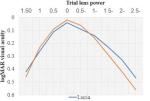
Figure 1 illustrates the postoperative defocus curves at 3 months. Defocus curve analysis of the PanOptix group demonstrated a broader range of functional near vision compared with the Vivity and Lucia groups, particularly at higher negative defocus levels, consistent with the trifocal optical design of the lens. At a visual acuity threshold of 0.2 logMAR, the defocus range of the Vivity group exceeded that of the Lucia group by 0.25 diopters. The mean intermediate visual acuity of the Vivity group was 0.238 logMAR, compared with 0.306 logMAR in the Lucia group, with a statistically significant difference (p = 0.035). The median intermediate visual acuity of the Vivity group was 0.2 logMAR. Distance visual acuity between the two groups was similar, with no statistically significant difference.
Table 3 reports the assessment of the ANSI extended depth-of-focus criteria at 3 months. The criterion regarding mean monocular intermediate visual acuity being better than that of the monofocal control lens was met, with mean values of 0.238 in the Vivity group and 0.306 in the Lucia group. The criterion for median monocular intermediate visual acuity of at least 0.2 logMAR was also met in the Vivity group. In addition, the criterion requiring mean distance-corrected visual acuity not to be worse than the monofocal control lens was fulfilled, with mean values of 0.042 in the Vivity group and 0.026 in the Lucia group.
Table 4 summarises spectacle dependence, visual disturbances, contrast sensitivity, and patient-reported outcomes at 3 months postoperatively. Most patients in the Vivity and PanOptix groups did not require spectacles, accounting for 90.0% and 94.0%, respectively, compared with 22.0% in the Lucia group. Visual disturbances such as halos, glare, or starbursts were reported in 16.0% of the Vivity group, 34.0% of the PanOptix group, and 16.0% of the Lucia group, with most cases classified as mild. Mean contrast sensitivity values at spatial frequencies of 1.5, 3.0, and 6.0 cycles per degree were recorded for all groups. At 3 months, the proportion of patients classified as very satisfied was 94.0% in the Vivity group, 76.0% in the PanOptix group, and 40.0% in the Lucia group. The mean VF-14 total score was 98.593 ± 2.549 for the Vivity group, 96.284 ± 3.828 for the PanOptix group, and 91.355 ± 5.281 for the Lucia group.
This study demonstrated that implantation of the non-diffractive extended depth-of-focus intraocular lens AcrySof IQ Vivity provided a balanced visual performance across distance and intermediate ranges, with visual acuity outcomes comparable to a diffractive multifocal intraocular lens and superior to a monofocal intraocular lens at intermediate distances. The Vivity lens fulfilled key ANSI criteria for extended depth-of-focus performance, showed high levels of spectacle independence, preserved contrast sensitivity, and was associated with a lower incidence of visual disturbances compared with the multifocal lens. Patient-reported satisfaction and functional vision scores were also high in the Vivity group.
At three months postoperatively, distance visual acuity in the Vivity group was comparable to that achieved with both the monofocal Lucia and the multifocal PanOptix intraocular lenses, confirming that the non-diffractive EDOF design does not compromise distance vision. Concerning near and intermediate visual performance, the Vivity lens provided better outcomes than the monofocal lens, while showing intermediate characteristics between the monofocal and multifocal designs. Near vision with Vivity was inferior to PanOptix but superior to Lucia, whereas intermediate vision was similar to PanOptix and better than Lucia. This pattern reflects the intended optical profile of the Vivity lens, which prioritises extending depth of focus into the intermediate range while maintaining high-quality distance vision. These findings are consistent with the multicenter, randomised, double-masked clinical trial by Bala et al., which evaluated the effectiveness and safety of the Vivity intraocular lens in comparison with a monofocal IOL (IQ). In that study, conducted across 19 sites in four countries with 282 patients, distance visual acuity after Vivity implantation was equivalent to that of the monofocal lens, while intermediate and near visual acuity were significantly better in the Vivity group [15]. Similarly, Won et al. reported that distance vision with Vivity was comparable to both monofocal and PanOptix lenses, intermediate vision was superior to monofocal and equivalent to PanOptix, and near vision remained better with PanOptix than with Vivity [16]. Comparable results were also observed in the randomised, double-masked study by McCabe et al., in which Vivity provided distance vision equivalent to a monofocal lens and superior intermediate and near visual performance at six months [17].
A high level of spectacle independence was observed in the Vivity group in the present study, with more than four-fifths of eyes not requiring spectacles for daily activities at different viewing distances. This finding aligns with previous studies reporting spectacle independence rates ranging from 70% to over 85% following Vivity implantation [15,17-19]. In contrast, higher spectacle independence associated with PanOptix has also been reported, as shown by Hovanesian et al. where multifocal lens implantation resulted in the highest rate of complete spectacle freedom, exceeding that of Vivity [20]. These findings underscore the trade-off between near vision performance and optical side effects among different IOL designs.
Regarding classification as an extended depth-of-focus intraocular lens, our results differ from those of Bala et al. [15] and McCabe et al. [17], both of whom concluded that Vivity fulfilled all ANSI criteria for EDOF lenses. This discrepancy may be attributed to differences in the monofocal control lens used for comparison. Whereas previous studies compared Vivity against the IQ monofocal lens, the present study used the Lucia monofocal lens as the control, which may have influenced the measured depth-of-focus threshold under ANSI standards.
Visual quality outcomes further support the advantages of the non-diffractive Vivity design. Most patients in the Vivity group reported no visual disturbances, with the remainder experiencing only mild symptoms, a profile comparable to the monofocal group and markedly better than the PanOptix group. These findings are in line with those of Monaco et al. who reported that more than 70% of patients implanted with Vivity experienced no halos or glare, with rates similar to those in monofocal lens recipients [21]. Comparable results have also been reported by Hovanesian et al. with a lower rate of visual disturbances in the Vivity group compared with PanOptix [20], as well as in other studies demonstrating similar disturbance profiles between Vivity and monofocal lenses [22-24].
Contrast sensitivity was well preserved in the Vivity group across low, medium, and high spatial frequencies and was comparable to the monofocal lens while exceeding that of the PanOptix lens. This finding is consistent with the study by Won et al. in which Vivity demonstrated contrast sensitivity similar to monofocal lenses and superior to PanOptix [16]. Previous studies have also reported reduced contrast sensitivity with diffractive multifocal lenses compared with monofocal optics, supporting the observed differences in this study [14,25].
Finally, patient-reported satisfaction was notably high in the Vivity group, as reflected by both subjective satisfaction ratings and VF-14 functional vision scores. The proportion of patients reporting high satisfaction with Vivity was comparable to that reported by Bhasin et al. who found high postoperative satisfaction and willingness to choose the same lens for the fellow eye or recommend it to others [26]. Multiple studies have also documented VF-14 scores exceeding 90 following Vivity implantation, indicative of excellent functional visual outcomes and quality of life [27,28].
The findings of this study suggest that the AcrySof IQ Vivity intraocular lens is a valuable option for patients undergoing cataract surgery who desire reduced spectacle dependence, particularly for distance and intermediate activities, while maintaining high visual quality and minimising photic phenomena. The Vivity lens may be especially suitable for patients who are sensitive to halos and glare or who prioritise visual quality over maximal near vision.
This study has several limitations. The follow-up period was limited to three months and may not capture longer-term visual adaptation or late complications. An important consideration when interpreting the findings of this study is the non-randomised group allocation process. Intraocular lens selection was influenced by patient preference, visual expectations, and financial considerations, which may have contributed to baseline demographic and clinical differences among groups. Consequently, residual confounding and selection bias cannot be fully excluded and may have affected postoperative visual and patient-reported outcomes despite standardised surgical and follow-up protocols. Additionally, the study was conducted at a single centre with a relatively modest sample size, which may limit the generalizability of the findings.
The non-diffractive extended depth-of-focus intraocular lens Vivity demonstrated favourable distance and intermediate visual outcomes, high spectacle independence, preserved contrast sensitivity, and low levels of visual disturbances. Compared with monofocal and multifocal intraocular lenses, Vivity may represent a balanced optical solution for cataract patients seeking functional vision across multiple distances with high visual quality.
What is known about this topic
- Extended depth-of-focus (EDOF) intraocular lenses were developed to provide a continuous range of functional vision while preserving contrast sensitivity and reducing photic phenomena compared with diffractive multifocal intraocular lenses;
- The non-diffractive AcrySof IQ Vivity intraocular lens has demonstrated effective distance and intermediate vision with fewer visual disturbances than multifocal designs in international studies. Still, comparative clinical evidence in Vietnamese cataract populations remains limited;
What this study adds
- This controlled clinical study in Vietnamese cataract patients shows that the Vivity EDOF intraocular lens provides distance vision comparable to monofocal and multifocal lenses, with intermediate vision superior to monofocal and similar to multifocal designs, along with high spectacle independence;
- Vivity implantation was associated with better contrast sensitivity, fewer photic phenomena than the diffractive multifocal lens, and the highest patient-reported functional vision and satisfaction, supporting its role as a balanced option for modern cataract surgery in Vietnam.
The authors declare no competing interests.
Le Viet Cuong: study conception, patient recruitment, data collection, and manuscript drafting. Kim Bao Giang: principal investigator, methodological supervision, data interpretation, and critical revision of the manuscript. Tham Truong Khanh Van: clinical assessment, data verification, and manuscript editing. All authors read and approved the final manuscript.
The authors sincerely thank the leadership and ophthalmology staff of Friendship Hospital and Vietnam National Eye Hospital for their support in patient recruitment, clinical assessment, and follow-up examinations. We are also grateful to all participating patients for their cooperation and contribution to this study.
Table 1: baseline characteristics of the study participants
Table 2: postoperative logMAR visual acuity outcomes among the three study groups
Table 3: ANSI criteria assessment
Table 4: visual outcome classification, astigmatism, spectacle dependence, and satisfaction among study groups at 3 months postoperatively
Figure 1: postoperative defocus curve at 3 months
- Spalton D, Koch D. The constant evolution of cataract surgery. BMJ. 2000 Nov 25;321(7272):1304. PubMed | Google Scholar
- Ang R, Quinto MMS, Cruz E, Rivera M, Martinez G. Comparison of clinical outcomes between femtosecond laser-assisted versus conventional phacoemulsification. Eye Vis (Lond). 2018 Apr 23;5:8. PubMed | Google Scholar
- Luo Y, Li H, Chen W, Gao Y, Ye Z, Li Z. Active-fluidics versus gravity-fluidics system in phacoemulsification for age-related cataract (AGSPC): study protocol for a prospective, randomised, double-blind, controlled clinical trial. BMJ Open. 2022 Jan 20;12(1):e059062. PubMed | Google Scholar
- Nørregaard J, Bernth-Petersen P, Bellan L, Alonso J, Black C, Dunn E et al. Intraoperative clinical practice and risk of early complications after cataract extraction in the United States, Canada, Denmark, and Spain. Ophthalmology. 1999 Jan;106(1):42-8. PubMed | Google Scholar
- Kanellopoulos A, Asimellis G. Standard manual capsulorhexis / Ultrasound phacoemulsification compared to femtosecond laser-assisted capsulorhexis and lens fragmentation in clear cornea small incision cataract surgery. Eye Vis (Lond). 2016 Jul 29;3:20. PubMed | Google Scholar
- Malvankar-Mehta M, Iordanous Y, Chen Y, Wang W, Patel S, Costella J et al. iStent with Phacoemulsification versus Phacoemulsification Alone for Patients with Glaucoma and Cataract: A Meta-Analysis. PLoS One. 2015 Jul 6;10(7):e0131770. PubMed | Google Scholar
- Sato T. Eight-Chop Technique in Phacoemulsification Using Iris Hooks for Patients with Cataracts and Small Pupils. J Clin Med. 2024 Nov 30;13(23):7298. PubMed | Google Scholar
- Calderon I, Laroche D. Affordable Sinskey Hook Goniotomy and Cataract Surgery in Black and Afro-Latino Patients Diagnosed with Glaucoma: Retrospective Real-World One-Year Results. J Clin Med. 2025 May 8;14(10):3266. PubMed | Google Scholar
- Savini G, Galzignato A, Coutinho CP, Huang J, Wu Y, Barboni P et al. Influence of preoperative variables on the 3-month functional outcomes of the Vivity extended depth-of-focus intraocular lens: a prospective case series. Eye Vis (Lond). 2025 Feb 2;12(1):8. PubMed | Google Scholar
- Bhasin P, Sarkar D, Bhasin P, Dhanapal P, Ubhal G, Bhargava M. Visual performance and patient satisfaction with AcrySof® IQ Vivity® IOL: Experience from a tertiary care center in central India. Indian J Ophthalmol. 2024 Apr 1;72(4):554-557. PubMed | Google Scholar
- Koh K, Tuuminen R, Na DG, Jeon S. Patient-reported outcome measures after non-diffractive extended range of field IOL implantation: PureSee versus Vivity. Acta Ophthalmol. 2026 Jan 30. PubMed | Google Scholar
- Suzuki H, Ota Y, Hata S, Minami K, Bissen-Miyajima H. Binocular visual function of Japanese eyes with nondiffractive extended depth-of-focus intraocular lenses made of high water-content hydrophobic acrylic material. Jpn J Ophthalmol. 2026 Mar;70(2):304-309. PubMed | Google Scholar
- Ginsburg AP. Next generation contrast sensitivity testing. Functional assessment of low vision. 1996;77.
- Kohnen T, Klaproth OK, Bühren J. Effect of intraocular lens asphericity on quality of vision after cataract removal: an intraindividual comparison. Ophthalmology. 2009 Sep;116(9):1697-706. PubMed | Google Scholar
- Bala C, Poyales F, Guarro M, Mesa RR, Mearza A, Varma DK et al. Multicountry clinical outcomes of a new nondiffractive presbyopia-correcting IOL. J Cataract Refract Surg. 2022 Feb 1;48(2):136-143. PubMed | Google Scholar
- Won YK, Choi SH, Chung TY, Lim DH. Clinical Outcomes after Bilateral Implantation of a Wavefront-Shaping Extended Depth of Focus (EDOF) IOL with Mini-Monovision. J Clin Med. 2024 May 30;13(11):3225. PubMed | Google Scholar
- McCabe C, Berdahl J, Reiser H, Newsom TH, Cibik L, Koch D et al. Clinical outcomes in a U.S. registration study of a new EDOF intraocular lens with a nondiffractive design. J Cataract Refract Surg. 2022 Nov 1;48(11):1297-1304. PubMed | Google Scholar
- Kerr NM, Moshegov S, Lim S, Simos M. Visual Outcomes, Spectacle Independence, and Patient-Reported Satisfaction of the Vivity Extended Range of Vision Intraocular Lens in Patients with Early Glaucoma: An Observational Comparative Study. Clin Ophthalmol. 2023;17:1515-1523. PubMed | Google Scholar
- Howes F, Rementeria-Capelo LA, Poyales F, Borovik A, Khoramnia R, Perez-Vives C et al. Visual Performance and Patient Satisfaction of Bilaterally Implanted Extended Depth of Focus Intraocular Lens: Outcomes of a Multicenter Registry. J Refract Surg. 2025 Feb;41(2):e131-e143. PubMed | Google Scholar
- Hovanesian JA, Jones M, Allen Q. The Vivity Extended Range of Vision IOL vs the PanOptix Trifocal, ReStor 2.5 Active Focus and ReStor 3.0 Multifocal Lenses: A Comparison of Patient Satisfaction, Visual Disturbances, and Spectacle Independence. Clin Ophthalmol. 2022;16:145-152. PubMed | Google Scholar
- Monaco G, Gari M, Di Censo F, Poscia A, Ruggi G, Scialdone A. Visual performance after bilateral implantation of 2 new presbyopia-correcting intraocular lenses: Trifocal versus extended range of vision. J Cataract Refract Surg. 2017 Jun;43(6):737-747. PubMed | Google Scholar
- Guarro M, Sararols L, Londoño GJ, Goñi I, Vázquez M, Ruiz S et al. Visual disturbances produced after the implantation of 3 EDOF intraocular lenses vs 1 monofocal intraocular lens. J Cataract Refract Surg. 2022 Dec 1;48(12):1354-1359. PubMed | Google Scholar
- Sabur H, Unsal U. Visual outcomes of non-diffractive extended-depth-of-focus and enhanced monofocal intraocular lenses: A case-control study. Eur J Ophthalmol. 2023 Jan;33(1):262-268. PubMed | Google Scholar
- Zeng M, Liu Y, Liu X, Yuan Z, Luo L, Xia Y et al. Aberration and contrast sensitivity comparison of aspherical and monofocal and multifocal intraocular lens eyes. Clin Exp Ophthalmol. 2007 May-Jun;35(4):355-60. PubMed | Google Scholar
- Rocha KM, Chalita MR, Souza CE, Soriano ES, Freitas LL, Muccioli C et al. Postoperative wavefront analysis and contrast sensitivity of a multifocal apodized diffractive IOL (ReSTOR) and three monofocal IOLs. J Refract Surg. 2005 Nov-Dec;21(6):S808-12. PubMed | Google Scholar
- Bhasin P, Sarkar D, Bhasin P, Dhanapal PP, Ubhal GN, Bhargava M. Visual performance and patient satisfaction with AcrySof® IQ Vivity® IOL: Experience from a tertiary care center in central India. Indian J Ophthalmol. 2024 Apr 1;72(4):554-557. PubMed | Google Scholar
- Lee MW. Visual outcomes following bilateral implantation of a non-diffractive extended depth of focus toric intraocular lens using a mini-monovision approach. Int Ophthalmol. 2024 Jul 24;44(1):334. PubMed | Google Scholar
- Giannuzzi F, Carlà MM, Margollicci F, Di Stefano G, Molle A, Hu L et al. Functional outcomes and quality of life after AcrySof IQ Vivity intraocular lens implantation in a real-world study. Sci Rep. 2024 Sep 4;14(1):20620. PubMed | Google Scholar
Search
This article authors
On Pubmed
On Google Scholar
Citation [Download]
Navigate this article
Similar articles in
Key words
Tables and figures

This supplement
Article metrics
Recently from the PAMJ





Authors´ services
