
TNF-α-associated activation of Notch-1/Hes-1 signalling through ERK1/2 in rheumatoid arthritis synoviocytes
Nguyen Van Trieu
Corresponding author: Nguyen Van Trieu, Faculty of Medicine, Dai Nam University, Hanoi, Vietnam 
Received: 17 Feb 2026 - Accepted: 26 Jun 2026 - Published: 06 Jul 2026
Domain: Molecular Biology
Keywords: Notch-1/Hes-1 signalling, Rheumatoid arthritis, ERK1/2 pathway
Funding: This work received no specific grant from any funding agency in the public, commercial, or non-profit sectors.
This article is published as part of the supplement Innovations and Challenges in Global Health: A Multidisciplinary Perspective, commissioned by Young Researchers and Elite Club.
©Nguyen Van Trieu et al. Pan African Medical Journal (ISSN: 1937-8688). This is an Open Access article distributed under the terms of the Creative Commons Attribution International 4.0 License (https://creativecommons.org/licenses/by/4.0/), which permits unrestricted use, distribution, and reproduction in any medium, provided the original work is properly cited.
Cite this article: Nguyen Van Trieu et al. TNF-α-associated activation of Notch-1/Hes-1 signalling through ERK1/2 in rheumatoid arthritis synoviocytes. Pan African Medical Journal. 2026;54(1):12. [doi: 10.11604/pamj.supp.2026.54.1.51671]
Available online at: https://www.panafrican-med-journal.com//content/series/54/1/12/full
 PDF in process
 Research 
TNF-α-associated activation of Notch-1/Hes-1 signalling through ERK1/2 in rheumatoid arthritis synoviocytes
TNF-α-associated activation of Notch-1/Hes-1 signalling through ERK1/2 in rheumatoid arthritis synoviocytes
Nguyen Van Trieu1,&
&Corresponding author
Introduction: this pilot mechanistic study aimed to characterise the expression of the Notch-1/Hes-1 signalling axis in synoviocytes (Sc) from normal, osteoarthritis (OA), and rheumatoid arthritis (RA) subjects, and to investigate the role of tumour necrosis factor-alpha (TNF-α) and ERK1/2 signalling in its activation.
Methods: synovial tissues were obtained from patients with RA (n = 3), OA (n = 3), and normal controls (n = 2). Primary Sc cultures (passages 2-5) were analysed by immunocytochemistry, immunofluorescence, and Western blot to evaluate Notch-1 and Hes-1 expression in whole-cell lysates and nuclear extracts. TNF-α stimulation and pharmacological inhibition experiments were performed to investigate pathway involvement, while transient MEK1/2 transfection was used to assess ERK1/2-mediated effects. Quantitative comparisons were analysed using one-way ANOVA followed by post hoc testing, with p < 0.05 considered statistically significant.
Results: notch-1 and Hes-1 were constitutively expressed in Sc, with significantly higher nuclear localisation in RA Sc compared with OA and normal Sc (p < 0.001). TNF-α induced dose-dependent Notch-1 activation in OA and normal Sc, increasing both membrane-bound (~117 kDa) and nuclear (~63 kDa) forms and upregulating Hes-1 expression. ERK1/2 inhibition with PD98059 abrogated TNF-α-induced Notch-1 activation, whereas MEK1/2 overexpression enhanced nuclear NICD and Hes-1 expression. These findings support the involvement of the TNF-α/ERK1/2/Notch-1/Hes-1 signalling pathway in synoviocytes.
Conclusion: the Notch-1/Hes-1 signalling axis appears to be constitutively activated in RA synoviocytes and may be induced by TNF-α through ERK1/2-dependent signalling. Further studies with larger sample sizes and functional validation are required to clarify its role in RA pathogenesis.

Notch receptors are evolutionarily conserved transmembrane proteins that orchestrate key developmental processes. In mammals, four Notch genes (Notch 1-4) and five canonical ligands-Delta-like (DLL) 1, 3, and 4, and Jagged 1 and 2-have been characterised [1,2]. Activation of the pathway triggers proteolytic release of the Notch intracellular domain (NICD) [1,3-5], which translocates to the nucleus, associates with the DNA-binding factor CSL (RBP-Jκ), and regulates transcription of target genes [6]. Among these, Hes-1, a basic helix-loop-helix (bHLH) transcription factor, suppresses lineage-specific differentiation by antagonising other bHLH proteins [7-9]. Notch signalling has been shown to govern differentiation, proliferation, and apoptosis, and dysregulation has been implicated in diverse human diseases [1,3-5].
Rheumatoid arthritis (RA) is a chronic inflammatory disease characterised by aberrant synovial cell proliferation and differentiation, driven in part by proinflammatory cytokines. Tumour necrosis factor alpha (TNF-α) is recognised as a central mediator in RA pathogenesis [10], and crosstalk between cytokine networks and Notch signalling has been documented [11-14]. Previous studies have demonstrated the expression of Notch signalling components in rheumatoid synovium and synoviocytes, including Notch homologues and TNF-α-associated Notch activation in rheumatoid synovial fibroblasts [11,13,15-18]. Functional expression of the Notch-1/Hes-1 pathway has also been reported in muscle, hematopoietic, and neuronal cells [15-17]. These findings established the involvement of Notch signalling in synovial inflammatory responses; however, the expression profile of Hes-1 in human synoviocytes and the molecular basis linking TNF-α stimulation to ERK1/2-mediated Notch-1/Hes-1 activation remain incompletely characterised.
In this study, the expression of Notch-1 and Hes-1 in synoviocytes was examined, the role of TNF-α in activating this signalling axis was evaluated, and the underlying molecular mechanism was elucidated. Specifically, the present work extends previous observations by characterising Hes-1 expression in synoviocytes and investigating the contribution of ERK1/2 signalling to TNF-α-associated activation of Notch-1/Hes-1. These findings were intended to clarify the interface between inflammatory cytokine signalling and developmental pathways in RA, offering potential targets for therapeutic modulation.

Study design: this was an experimental laboratory-based pilot mechanistic study designed to characterise Notch-1/Hes-1 signalling and its regulation by tumour necrosis factor-alpha (TNF-α) and ERK1/2 pathways in primary human synoviocytes. The study combined comparative expression analysis across disease groups with cytokine stimulation, pharmacologic inhibition, and gain-of-function transfection experiments. Because this was an in vitro mechanistic study rather than an observational epidemiological study, STROBE reporting guidelines were not applied. Methodological reporting was structured according to principles of transparent bioscience reporting and MDAR-style recommendations for experimental studies.
Setting: synovial tissue specimens were obtained from patients undergoing orthopaedic surgery in a tertiary clinical setting. Laboratory experiments, including cell culture, immunofluorescence, Western blotting, inhibitor assays, and transfection experiments, were conducted in a controlled biomedical research laboratory using standardised protocols and reagents.
Human research participants and synovial tissue collection: baseline donor characteristics were recorded for all study participants. The RA donor group consisted of two females and one male, with a mean age of 57.3 ± 6.1 years and a median disease duration of 8 years (range: 5-12 years). All RA donors had previously received conventional disease-modifying antirheumatic drugs, including methotrexate and sulfasalazine, and one donor had prior biologic therapy exposure. The OA donor group consisted of two females and one male, with a mean age of 63.7 ± 5.4 years. None of the OA donors had a documented history of autoimmune inflammatory arthritis or biologic therapy exposure. Normal synovial tissues were obtained from two trauma patients without clinical evidence of inflammatory arthritis, autoimmune disease, or chronic synovitis. The normal donor group included one female and one male, with a mean age of 38.5 ± 4.9 years. All synovial tissues were collected before experimental manipulation. Tissue specimens were anonymised before laboratory processing.
Primary synoviocyte isolation and culture: primary synoviocytes were isolated by collagenase digestion of synovial tissue followed by adherence purification and expansion in Dulbecco's modified Eagle medium supplemented with 10% fetal calf serum. Cells were maintained under standard humidified culture conditions with 5% CO₂ at 37°C. Primary synoviocytes from passages two to five were used for all analyses to minimise contamination by non-adherent inflammatory cells while preserving primary synoviocyte characteristics. Experiments using later passages were avoided because of potential phenotypic drift during prolonged culture.
Experimental variables and pathway manipulation: the principal biological variables were the expression and nuclear localisation of Notch-1 intracellular domain (NICD) and Hes-1 in synoviocytes. Experimental exposure variables included TNF-α stimulation and pharmacologic inhibition of ERK1/2, p38, JNK, IKK/PKC, sphingomyelinase, and caspase-8 pathways. Gain-of-function variables included MEK1/2 overexpression. For pathway activation experiments, synoviocytes were exposed to recombinant TNF-α at specified concentrations for assay-dependent durations. TNF-α exposure duration was reported separately for immunocytochemistry, immunofluorescence, and Western blot analyses. For inhibition studies, synoviocytes were pretreated with PD98059 or other pathway inhibitors before TNF-α stimulation. Concentration and pretreatment duration for each inhibitor were predefined and maintained consistently within experiments. For gain-of-function studies, synoviocytes were transiently transfected with MEK1/2 expression plasmid using FuGENE6 transfection reagent. Transfection efficiency was monitored by GFP fluorescence and quantified as the percentage of GFP-positive cells.
Immunocytochemistry and immunofluorescence: immunocytochemistry and immunofluorescence staining involved paraformaldehyde fixation, Triton X-100 permeabilisation, incubation with primary antibodies, secondary antibody labelling, and fluorescence microscopy visualisation. Nuclear NICD expression was quantified by counting positive nuclei per 100 cells across five randomly selected ×400 microscopic fields per well. All antibodies used in the study, including supplier name, clone information, catalogue number, working dilution, and validation details.
Western blot analysis: protein expression was assessed by SDS-PAGE and Western blot analysis using specific primary antibodies against Notch-1 and Hes-1. Whole-cell lysates and nuclear extracts were prepared using standardised extraction procedures. Protein loading amount per lane, membrane type, blocking conditions, exposure method, exposure duration, and normalisation controls for whole-cell and nuclear fractions were predefined and maintained consistently across experiments. Chemiluminescent detection was performed according to the manufacturer's instructions.
Bias control and reproducibility: selection bias was minimised by using clinically defined RA, OA, and normal donor groups according to standardised diagnostic criteria. Measurement bias was reduced through blinded counting of NICD-positive nuclei across predefined microscopic fields. Technical reproducibility was strengthened through duplicate wells within each experimental condition, while biological reproducibility was assessed using independent donor-derived primary cultures. All experiments were independently repeated at least three times. Biological replicates referred to synoviocytes derived from independent human donors, whereas technical replicates referred to repeated measurements performed within the same experimental condition.
Study size and replication strategy: the experimental sample comprised synovial tissue from eight donors, including RA n = 3, OA n = 3, and normal n = 2. This work was designed as a pilot exploratory mechanistic study rather than a statistically powered clinical investigation. The limited number of donors, particularly within the normal control group, represented an important limitation affecting statistical power and generalizability. Within each experimental condition, assays were performed in duplicate wells and repeated in at least three independent experiments to support reproducibility of molecular findings.
Quantitative variables and statistical analysis: quantitative outcomes included the proportion of NICD-positive nuclei per 100 cells and densitometric protein expression levels derived from Western blot assays. Results were expressed as mean ± standard error of the mean (SEM). Individual biological data points were displayed where possible alongside summary statistics. For comparisons involving three disease groups, quantitative variables were analysed using one-way analysis of variance followed by Tukey post hoc testing. For comparisons involving two experimental conditions, an unpaired Student's t-test was used. When multiple pairwise comparisons were performed, the Bonferroni correction was applied. Statistical significance was defined as p < 0.05. The exact biological sample size, number of independent experiments, and number of technical replicates were specified for each analysis. Statistical analyses were performed using appropriate statistical software.
Ethics statement: all human synovial tissue samples were obtained with written informed consent from participants before surgery. The institutional ethics committee approved the study protocol in accordance with the Declaration of Helsinki. Tissue specimens were anonymised, and all procedures complied with ethical standards for research involving human biological material.
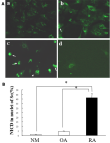
Expression of Notch-1 and Hes-1 in synoviocytes: the expression and subcellular localisation of Notch-1 in synoviocytes (Sc) were first examined by immunofluorescence and Western blot analysis. Notch-1 expression was detected in normal, osteoarthritis (OA), and rheumatoid arthritis (RA) synoviocytes, with markedly greater nuclear localisation observed in RA Sc. No staining was detected in isotype antibody controls. Representative immunofluorescence images showing NICD expression in synoviocytes from normal (a), OA (b), and RA (c) donors. Panel (d) represents the isotype antibody control. Green fluorescence indicates NICD expression, and nuclei were counterstained with DAPI. Arrows indicate representative NICD-positive nuclei. Images were acquired using identical exposure and gain settings. Scale bar = 50 μm (Figure 1A). Quantitative analysis demonstrated that NICD-positive nuclei accounted for only 1.1 ± 0.33% of cells in normal Sc and 4.1 ± 0.8% in OA Sc, compared with 41.2 ± 4.5% in RA Sc (p < 0.001). Quantification of NICD-positive nuclei expressed as the percentage of NICD-positive nuclei. Quantification was performed by blinded counting of 100 cells across five randomly selected ×400 microscopic fields per well using independent biological donor-derived cultures (normal n = 2, OA n = 3, RA n = 3), with duplicate technical replicates for each condition. Data are presented as mean ± SEM. Statistical comparisons between groups were performed using unpaired two-tailed Student's t-tests. ***p < 0.001 versus the RA group (Figure 1B).
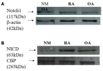
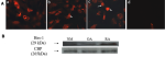
Western blot analysis of whole-cell lysates demonstrated comparable expression of membrane-associated Notch-1 (~117 kDa) among normal, OA, and RA synoviocytes (Figure 2A). In contrast, Western blot analysis of nuclear extracts revealed markedly increased nuclear accumulation of the ~63-kDa Notch intracellular domain (NICD) in RA Sc relative to normal and OA Sc (Figure 2B), indicating enhanced constitutive Notch-1 activation in RA synoviocytes. Because Hes-1 is a major downstream transcriptional target of Notch-1 signalling [1,6], its expression was subsequently evaluated. Immunofluorescence analysis demonstrated nuclear Hes-1 expression in all synoviocyte groups, with substantially greater nuclear accumulation in RA Sc compared with OA and normal Sc. Representative immunofluorescence images showing Hes-1 expression in synoviocytes from normal (a), OA (b), and RA (c) donors. Panel (d) represents the isotype antibody control. Red fluorescence indicates Hes-1 expression, and arrows indicate representative Hes-1-positive nuclei. Images were acquired using identical exposure and gain settings under ×400 magnification. Scale bar = 50 μm (Figure 3A). Consistently, Western blot analysis of nuclear extracts showed increased Hes-1 protein expression (~29 kDa) in RA synoviocytes relative to OA and normal controls. Representative Western blot analysis of nuclear extracts demonstrating Hes-1 expression (~29 kDa) in synoviocytes derived from normal (NM), OA, and RA donors. CBP (~265 kDa) was used as the nuclear loading control. Lane assignment was as follows: lane 1 = NM, lane 2 = OA, and lane 3 = RA. Representative blots were selected from at least three independent experiments performed using donor-derived biological replicates under identical exposure conditions. Densitometric quantification of Hes-1 normalized to CBP (Figure 3B). Collectively, these findings indicate constitutive activation of the Notch-1/Hes-1 signaling axis in RA synoviocytes.
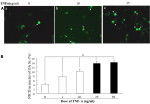
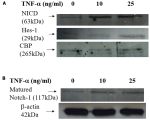
TNF-α activation of Notch-1 and Hes-1 in synoviocytes: given the established role of TNF-α in synoviocyte activation and proliferation, its involvement in regulating Notch-1 signalling was investigated. Immunofluorescence analysis demonstrated that TNF-α induced dose-dependent nuclear translocation of NICD in OA synoviocytes. Representative immunofluorescence images of OA synoviocytes following TNF-α stimulation at 0 ng/ml (a), 10 ng/ml (b), and 25 ng/ml (c) for 4 h, demonstrating dose-dependent nuclear accumulation of NICD. Green fluorescence indicates NICD expression, and arrows indicate representative NICD-positive nuclei. Images were acquired using identical exposure and gain settings under ×400 magnification. Scale bar = 50 μm. (Figure 4A). Quantification showed that NICD-positive nuclei increased from approximately 4% in untreated cells to approximately 14% following TNF-α stimulation at 50 ng/ml. Quantification of NICD-positive nuclei expressed as the percentage of NICD-positive nuclei following TNF-α stimulation (0, 1, 10, 25, and 50 ng/ml). Nuclear NICD-positive cells increased from approximately 4% in untreated OA synoviocytes to approximately 14% following TNF-α treatment at 50 ng/ml. Quantification was performed by blinded counting of 100 cells across five randomly selected ×400 microscopic fields per well using independent biological donor-derived OA synoviocyte cultures, with duplicate technical replicates for each condition. Data are presented as mean ± SEM. Statistical comparisons between TNF-α-treated and untreated groups were performed using unpaired two-tailed Student's t-tests. *p < 0.01 versus untreated control (0 ng/ml) (Figure 4B). A similar induction pattern was observed in normal synoviocytes, whereas only minimal additional activation was detected in RA Sc, likely reflecting their high baseline NICD activity. To further characterise TNF-α-mediated signalling, Western blot analyses were performed. Nuclear extract analysis demonstrated dose-dependent increases in both NICD and Hes-1 protein expression following TNF-α stimulation in OA synoviocytes. Representative Western blot analysis of nuclear extracts from OA synoviocytes following TNF-α stimulation (0, 10, and 25 ng/ml for 4 h). TNF-α induced dose-dependent increases in nuclear Notch intracellular domain (NICD; ~63 kDa) and Hes-1 (~29 kDa) expression. CBP (~265 kDa) was used as the nuclear loading control. (Figure 5A). In parallel, whole-cell lysate analysis showed increased expression of mature membrane-associated Notch-1 (~117 kDa) after TNF-α treatment. Representative Western blot analysis of whole-cell lysates from OA synoviocytes following TNF-α stimulation (0, 10, and 25 ng/ml for 4 h). TNF-α increased the expression of mature Notch-1 (~117 kDa) in a dose-dependent manner. β-actin (~42 kDa) was used as the loading control. (Figure 5B). Similar TNF-α-induced Hes-1 upregulation was also observed in normal synoviocytes (data not shown). These findings suggest that TNF-α promotes activation of the Notch-1/Hes-1 signalling pathway in synoviocytes.
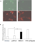
ERK1/2-mediated induction of Notch-1 and Hes-1: to investigate the signalling mechanism underlying TNF-α-induced Notch-1 activation, both pharmacologic inhibition and gain-of-function approaches were employed. Pretreatment with PD98059, a selective ERK1/2 inhibitor, completely abrogated TNF-α-induced NICD nuclear translocation in OA synoviocytes, reducing NICD-positive nuclei to near-baseline levels (5.9 ± 0.6% vs. 4.2 ± 0.5%; p > 0.05). In contrast, inhibitors targeting p38, sphingomyelinase, IKK, PKC, caspase-8, or JNK pathways exerted minimal or no inhibitory effects (data not shown). Western blot analysis further confirmed that PD98059 selectively suppressed TNF-α-induced Notch-1 expression in OA synoviocytes. To further validate the involvement of ERK1/2 signalling, OA synoviocytes were transiently transfected with a MEK1/2 expression plasmid (MEK1/2-GFP). Immunofluorescence analysis demonstrated increased nuclear NICD accumulation in MEK1/2-transfected cells compared with empty vector controls. Representative immunofluorescence images of OA synoviocytes transfected with MEK1/2-GFP expression plasmid or empty vector control. Panels (a) and (b) show GFP fluorescence indicating transfection efficiency, whereas panels (c) and (d) show NICD immunofluorescence. Arrows indicate representative NICD-positive nuclei. MEK1/2 overexpression increased nuclear NICD accumulation compared with empty vector control. Images were acquired using identical exposure and gain settings under ×400 magnification. Scale bar = 50 μm (Figure 6A). Quantitative analysis revealed an approximately two-fold increase in NICD-positive nuclei following MEK1/2 overexpression (10 ± 0.5% vs. 5 ± 0.9%; p < 0.001). Quantification of NICD-positive nuclei expressed as the percentage of NICD-positive nuclei in OA synoviocytes under medium control, empty vector (pcDNA3), MEK1/2 overexpression, and TNF-α stimulation (25 ng/ml) conditions. Quantification was performed by blinded counting of 100 cells across five randomly selected ×400 microscopic fields per well using independent biological donor-derived cultures, with duplicate technical replicates for each condition. Data are presented as mean ± SEM. Transfection efficiency was monitored by GFP fluorescence before analysis. Statistical comparisons between groups were performed using unpaired two-tailed Student's t-tests. p < 0.01 versus pcDNA3 control (Figure 6B). Western blot analysis of nuclear extracts further confirmed increased NICD expression after MEK1/2 overexpression. Because Hes-1 functions downstream of Notch-1 activation, its regulation by ERK1/2 signalling was also examined. MEK1/2 overexpression significantly increased nuclear Hes-1 protein expression in OA synoviocytes, whereas no comparable increase was observed in empty vector-transfected controls. Immunofluorescence analysis further demonstrated marked nuclear accumulation and co-localisation of Hes-1 in MEK1/2-transfected cells (data not shown). Together, these findings support the involvement of the TNF-α/ERK1/2/Notch-1/Hes-1 signalling pathway in synoviocyte activation.
The functional expression of Notch receptors and their downstream effector Hes-1 has been documented in multiple cell types, including neurons, myocytes, and hematopoietic cells. Previous work demonstrated that Notch-1, Notch-2, and Notch-3 are expressed in synoviocytes (Sc) [18], although the expression pattern of Hes-1 in these cells had not been well characterised. The present findings extend these previous observations by demonstrating constitutive expression of both Notch-1 and Hes-1 in synoviocytes and by showing increased nuclear localisation of these proteins in rheumatoid arthritis (RA) synoviocytes compared with osteoarthritis (OA) and normal controls. Nuclear NICD levels were approximately 10-fold greater than OA and 30-fold greater than normal controls. As nuclear localisation is a hallmark of Notch activation [6,8,19-21], these findings suggest that Notch-1 signalling is constitutively activated in RA synoviocytes.
TNF-α was shown to induce Notch-1 expression in OA and normal Sc in a dose-dependent manner, promoting nuclear NICD accumulation as confirmed by immunofluorescence. Western blot analysis demonstrated increases in both membrane-bound (~117 kDa) and nuclear (~63 kDa) NICD following TNF-α stimulation, consistent with its role as a potent regulator of Notch signalling [22-24]. Although nuclear NICD-positive cells did not exceed 20% of the total population, prior evidence suggests that relatively small amounts of nuclear NICD may be sufficient for transcriptional activity [25]. These findings are consistent with previous reports identifying TNF-α as an activator of Notch signalling in synovial cells [13,14,18,26], and with studies demonstrating that Notch activation may contribute to proinflammatory cytokine production in RA [27-30]. Collectively, these observations support the possibility that TNF-α-mediated Notch activation may contribute to synovial inflammation and activation in RA.
Mechanistically, TNF-α-induced Notch-1 activation appeared to be mediated predominantly through the ERK1/2 MAPK pathway. PD98059, a selective ERK1/2 inhibitor, abrogated NICD nuclear translocation, whereas inhibitors of p38, sphingomyelinase, IKK, PKC, caspase-8, or JNK exerted little or no effect. Gain-of-function studies using MEK1/2 overexpression enhanced Notch-1 and Hes-1 expression, providing mechanistic evidence supporting involvement of the TNF-α/ERK1/2/Notch-1/Hes-1 signalling axis in synoviocytes. Given the established roles of TNF-α, ERK1/2, and Notch-1 in RA [31-42], these findings suggest potential signalling cross-talk that may contribute to amplification of inflammatory responses in RA synoviocytes.
Hes proteins are basic helix-loop-helix transcription factors that regulate lineage determination and cell differentiation [7-9]. Among these, Hes-1 is a well-recognised Notch target gene [9] and, in this study, was found to be upregulated in RA Sc and positively regulated by TNF-α via ERK1/2. This coordinated upregulation supports the possibility that Hes-1 functions downstream of Notch-1 activation in synoviocytes. Functionally, Hes-1 is known to inhibit differentiation and participate in diverse developmental processes, including neurogenesis, myogenesis, hematopoiesis, and T-cell maturation [11,22,43-45]. Inflammatory relevance is underscored by its reported association with IL-6, IL-1 receptor-like 1, and MMP13 expression [9], and by evidence that inhibition of the Notch-Hes-1 axis ameliorates experimental arthritis [14,46,47]. However, the present study did not directly evaluate the effects of Notch-1/Hes-1 signalling on synoviocyte proliferation, migration, cytokine production, or matrix metalloproteinase expression, and therefore, the downstream functional significance of this pathway in RA remains incompletely defined.
Nonetheless, the role of Hes-1 in Notch-1 signalling within Sc may not be exclusive. Some studies have reported minimal Hes induction following stimulation with Delta-like 1 or Jagged-1 [23,48], and Notch signalling can proceed through Hes-independent pathways [23,35]. Conversely, Hes-1 may function independently of Notch-1 in certain contexts [35]. Thus, while the present results strongly support a Notch-1-associated mechanism for Hes-1 regulation in Sc, the possibility of alternative or parallel pathways cannot be excluded.
Several limitations of this study should be acknowledged. First, the donor sample size was small, particularly for the normal control group (n = 2), which limits statistical power and generalizability. Second, this was an in vitro mechanistic study using primary synoviocyte cultures, and therefore the findings may not fully reflect the complexity of the in vivo synovial microenvironment. Third, functional assays evaluating proliferation, migration, cytokine secretion, or matrix remodelling activity were not performed. Finally, in vivo validation studies were not conducted, and additional experimental and translational studies are required to confirm the biological and clinical relevance of these observations.
In summary, the present study extends previous observations of Notch signalling in synoviocytes by demonstrating constitutive activation of the Notch-1/Hes-1 axis in RA synoviocytes and by providing mechanistic evidence supporting TNF-α-induced activation through an ERK1/2-dependent pathway. These findings suggest that Notch-1/Hes-1 signalling may represent a mechanistic link between inflammatory cytokine signalling and synoviocyte activation in RA. Although the precise downstream functional consequences of this pathway remain to be clarified, the results support the possibility that Notch-1/Hes-1 signalling may contribute to inflammatory and proliferative responses in RA synoviocytes. Further in vitro and in vivo studies are needed to determine the functional significance of this pathway and to evaluate whether modulation of Notch signalling may represent a potential therapeutic target in RA and other chronic inflammatory conditions.
What is known about this topic
- Notch signalling regulates cell differentiation, proliferation, and inflammatory responses, and previous studies have demonstrated expression and activation of Notch pathway components in rheumatoid arthritis synoviocytes;
- Tumour necrosis factor-alpha (TNF-α) is a central cytokine in rheumatoid arthritis and is known to interact with intracellular signalling pathways, including MAPK signalling, although the mechanisms linking TNF-α to downstream Notch-1/Hes-1 activation in synoviocytes remain incompletely understood.
What this study adds
- This study demonstrates increased nuclear localisation of NICD and Hes-1 in rheumatoid arthritis synoviocytes compared with osteoarthritis and normal synoviocytes, supporting constitutive activation of the Notch-1/Hes-1 signalling axis in rheumatoid arthritis;
- The findings provide mechanistic evidence that TNF-α-induced Notch-1 activation in synoviocytes may occur through an ERK1/2-dependent pathway, as supported by pharmacologic inhibition and MEK1/2 overexpression experiments;
- These results extend previous observations of Notch signalling in rheumatoid arthritis and support the possibility that the TNF-α/ERK1/2/Notch-1/Hes-1 axis may contribute to synoviocyte activation and inflammatory responses.
The author declares no competing interests.
The author read and approved the final version of the manuscript.
The authors sincerely thank the leadership, surgeons, anesthesiologists, nurses, and medical staff of Nghe An Friendship General Hospital for their support in patient care, follow-up, and data collection.
Figure 1A, B): nuclear localisation of Notch-1 intracellular domain (NICD) in synoviocytes derived from normal, osteoarthritis (OA), and rheumatoid arthritis (RA)
Figure 2: membrane and nuclear Notch-1 in synoviocytes by Western blot: A) whole-cell lysates show ~117-kDa membrane Notch-1 in normal, RA, and OA synoviocytes; β-actin control; B) nuclear extracts show ~63-kDa NICD; CBP control
Figure 3A, B): nuclear expression of Hes-1 in synoviocytes derived from normal, osteoarthritis (OA), and rheumatoid arthritis (RA) donors
Figure 4A, B): TNF-α induces dose-dependent nuclear accumulation of Notch-1 intracellular domain (NICD) in osteoarthritis (OA) synoviocytes
Figure 5A, B): TNF-α regulates Notch-1 and Hes-1 expression in osteoarthritis (OA) synoviocytes
Figure 6A, B): MEK1/2 overexpression promotes nuclear accumulation of Notch-1 intracellular domain (NICD) in osteoarthritis (OA) synoviocytes
- Artavanis-Tsakonas S, Rand MD, Lake RJ. Notch signaling: cell fate control and signal integration in development. Science. 1999 Apr 30;284(5415):770-6. PubMed | Google Scholar
- Lai EC. Notch signaling: control of cell communication and cell fate. Development. 2004 Mar;131(5):965-73. PubMed | Google Scholar
- Gao W, Sweeney C, Walsh C, Rooney P, McCormick J, Veale DJ et al. Notch signalling pathways mediate synovial angiogenesis in response to vascular endothelial growth factor and angiopoietin 2. Ann Rheum Dis. 2013 Jun;72(6):1080-8. PubMed | Google Scholar
- Keewan E, Naser SA. The role of notch signaling in macrophages during inflammation and infection: implication in rheumatoid arthritis? Cells. 2020 Jan 2;9(1):111. PubMed | Google Scholar
- Ren H, Mu C, Wang Y, Cheng Y, Hou Y, Li Y et al. Notch2 Inhibition and Kidney Cyst Growth in Autosomal Dominant Polycystic Kidney Disease. J Am Soc Nephrol. 2025 May 1;36(5):781-797. PubMed | Google Scholar
- Jarriault S, Brou C, Logeat F, Schroeter EH, Kopan R, Israel A. Signaling downstream of activated mammalian Notch. Nature. 1995 Sep 28;377(6547):355-8. PubMed | Google Scholar
- Zhou B, Lin W, Long Y, Yang Y, Zhang H, Wu K et al. Notch signaling pathway: architecture, disease, and therapeutics. Signal Transduct Target Ther. 2022 Mar 24;7(1):95. PubMed | Google Scholar
- Artavanis-Tsakonas S, Matsuno K, Fortini ME. Notch signaling. Science. 1995 Apr 14;268(5208):225-32. PubMed | Google Scholar
- Sugita S, Hosaka Y, Okada K, Mori D, Yano F, Kobayashi H et al. Transcription factor Hes1 modulates osteoarthritis development in cooperation with calcium/calmodulin-dependent protein kinase 2. Proc Natl Acad Sci U S A. 2015 Mar 10;112(10):3080-5. PubMed | Google Scholar
- Mateen S, Zafar A, Moin S, Khan AQ, Zubair S. Understanding the role of cytokines in the pathogenesis of rheumatoid arthritis. Clin Chim Acta. 2016 Apr 1;455:161-71. PubMed | Google Scholar
- Ando K, Kanazawa S, Tetsuka T, Ohta S, Jiang X, Tada T et al. Induction of Notch signaling by tumor necrosis factor in rheumatoid synovial fibroblasts. Oncogene. 2003 Oct 30;22(49):7796-803. PubMed | Google Scholar
- Lu Y, Zhang Y, Xiang X, Sharma M, Liu K, Wei J et al. Notch signaling contributes to the expression of inflammatory cytokines induced by highly pathogenic porcine reproductive and respiratory syndrome virus infection in porcine alveolar macrophages. Dev Comp Immunol. 2020 Jul;108:103690. PubMed | Google Scholar
- Ishii H, Nakazawa M, Yoshino S, Nakamura H, Nishioka K, Nakajima T. Expression of notch homologues in the synovium of rheumatoid arthritis and osteoarthritis patients. Rheumatol Int. 2001 Sep;21(1):10-4. PubMed | Google Scholar
- Park JS, Kim SH, Kim K, Jin CH, Choi KY, Jang J et al. Inhibition of notch signalling ameliorates experimental inflammatory arthritis. Ann Rheum Dis. 2015 Jan;74(1):267-74. PubMed | Google Scholar
- Bjornson CR, Cheung TH, Liu L, Tripathi PV, Steeper KM, Rando TA. Notch signaling is necessary to maintain quiescence in adult muscle stem cells. Stem Cells. 2012 Feb;30(2):232-42. PubMed | Google Scholar
- Bigas A, Martin DIK, Milner L. Notch1 and Notch2 inhibit myeloid differentiation in response to different cytokines. Mol Cell Biol. 1998 Apr;18(4):2324-33. PubMed | Google Scholar
- Zhu J, Liu Q, Jiang Y, Wu L, Xu G, Liu X. Enhanced angiogenesis promoted by human umbilical mesenchymal stem cell transplantation in stroked mouse is Notch1 signaling associated. Neuroscience. 2015; 290:288-297. PubMed | Google Scholar
- Nakazawa M, Ishii H, Aono H, Takai M, Honda T, Aratani S et al. Role of Notch1 intracellular domain in activation of rheumatoid synoviocytes. Arthritis Rheum. 2001 Jul;44(7):1545-54. PubMed | Google Scholar
- Arnett FC, Edworthy SM, Bloch DA, McShane DJ, Pries JF, Cooper NS et al. The American Rheumatism Association 1987 revised criteria for the classification of rheumatoid arthritis. Arthritis Rheum. 1988 Mar;31(3):315-24. PubMed | Google Scholar
- Altman R, Alarcon G, Appelrouth D, Bloch D, Borenstein D, Brandt K et al. The American College of Rheumatology criteria for the classification and reporting of osteoarthritis of the hip. Arthritis Rheum. 1991 May;34(5):505-14. PubMed | Google Scholar
- Milner LA, Bigas A, Kopan R, Brashemstein C, Bernstein ID, Martin ID. Inhibition of granulocytic differentiation by mNotch1. Proc Natl Acad Sci U S A. 1996 Nov 12;93(23):13014-9. PubMed | Google Scholar
- Ohtsuka T, Ishibashi M, Gradwohl G, Nakanishi S, Guillemot F, Kageyama R. Hes1 and Hes5 as Notch effectors in mammalian neuronal differentiation. EMBO J. 1999 Apr 15;18(8):2196-207. PubMed | Google Scholar
- Shawber C, Nofziger D, Hsieh JJD, Lindsell C, Bögler O, Hayward D et al. Notch signaling inhibits muscle differentiation through a CBF1-independent pathway. Development. 1996 Dec;122(12):3765-73. PubMed | Google Scholar
- Fortini ME, Rebay I, Caron LA, Artavanis-Tsakonas S. An activated Notch receptor blocks cell-fate commitment in the developing drosophila eye. Nature. 1993 Oct 7;365(6446):555-7. PubMed | Google Scholar
- Varnum-Finney B, Brashemstein B, Bernstein ID. Combined effects of Notch signaling and cytokines induce a multiple log increase in precursors with lymphoid and myeloid reconstituting ability. Blood. 2003 Mar 1;101(5):1784-9. PubMed | Google Scholar
- Chen J, Li J, Chen J, Cheng W, Lin J, Ke L et al. Treatment of collagen-induced arthritis rat model by using Notch signalling inhibitor. J Orthop Translat. 2021 Mar 17;28:100-107. PubMed | Google Scholar
- Fung E, Tang SM, Canner JP, Morishige K, Arboleda-Velasquez JF, Cardoso AA et al. Delta-like 4 induces notch signaling in macrophages: implications for inflammation. Circulation. 2007 Jun 12;115(23):2948-56. PubMed | Google Scholar
- Jiao Z, Wang W, Ma J, Wang S, Su Z, Xu H. Notch signaling mediates TNF-α-induced IL-6 production in cultured fibroblast-like synoviocytes from rheumatoid arthritis. Clin Dev Immunol. 2012;2012:350209. PubMed | Google Scholar
- Su J, Tao Y, Liu J, Sun J, Zeng Y, Meng X et al. Tibetan medicine Qi-Sai-Er-Sang-Dang-Song decoction inhibits TNF-alpha-induced rheumatoid arthritis in human fibroblast-like synoviocytes via regulating NOTCH1/NF-kappaB/NLRP3 pathway. J Ethnopharmacol. 2023 Jun 28;310:116402. PubMed | Google Scholar
- Zack SR, Meyer A, Zanotti B, Volin MV, Deen S, Satoeya N et al. Notch ligands are biomarkers of anti-TNF response in RA patients. Angiogenesis. 2024 May;27(2):273-283. PubMed | Google Scholar
- Sujitha S, Rasool M. MicroRNAs and bioactive compounds on TLR/MAPK signaling in rheumatoid arthritis. Clin Chim Acta. 2017; 473:106-115. PubMed | Google Scholar
- Kanai T, Kondo N, Okada M, Sano H, Okumura G, Kijima Y et al. The JNK pathway represents a novel target in the treatment of rheumatoid arthritis through the suppression of MMP-3. J Orthop Surg Res. 2020 Mar 4;15(1):87. PubMed | Google Scholar
- Mittal S, Sharma A, Balaji SA, Gowda MC, Dighe RR, Kumar RV et al. Coordinate hyperactivation of notch1 and Ras/MAPK pathways correlates with poor patient survival: novel therapeutic strategy for aggressive breast cancers. Mol Cancer Ther. 2014 Dec;13(12):3198-3209. PubMed | Google Scholar
- Porcelli L, Mazzotta A, Garofoli M, DiFonte R, Guida G, Guida M et al. Active notch protects MAPK activated melanoma cell lines from MEK inhibitor cobimetinib. Biomed Pharmacother. 2021 Jan;133:111006. PubMed | Google Scholar
- Sriuranpong V, Borges MW, Rajani K, Ravi R, Arnold DR, Nelkin BD et al. Notch signaling induces cell cycle arrest in small cell lung cancer cells. Cancer Res. 2001 Apr 1;61(7):3200-5. PubMed | Google Scholar
- Nakajawa M, Ishii H, Nakamura H, Yoshino S, Fukamizu A, Nishioka K et al. NFkappaB2 (p52) promoter activation via Notch signaling pathway in rheumatoid arthritis. Int J Mol Med. 2001; 7:31-35. PubMed | Google Scholar
- Tak PP, Firestein GS. NF-κB: a key role in inflammatory diseases. J Clin Invest. 2001; 107(1):7-11. PubMed | Google Scholar
- Bash J, Zong WX, Banga S, Rivera A, Ballard DW, Ron Y et al. Rel/NF-κB can trigger the Notch signaling pathway by inducing the expression of Jagged1, a ligand for Notch receptors. EMBO J. 1999 May 17;18(10):2803-11. PubMed | Google Scholar
- Sato C, Zhao G, Ilagan MX. An overview of notch signaling in adult tissue renewal and maintenance. Curr Alzheimer Res. 2012 Feb;9(2):227-40. PubMed | Google Scholar
- Gao W, Sweeney C, Connolly M, Kennedy A, Ng CT, McCormick J et al. Notch-1 mediates hypoxia-induced angiogenesis in rheumatoid arthritis. Arthritis Rheum. 2012 Jul;64(7):2104-13. PubMed | Google Scholar
- Chen J, Cheng W, Li J, Wang Y, Chen J, Shen X et al. Notch-1 and Notch-3 mediate hypoxia-induced activation of synovial fibroblasts in rheumatoid arthritis. Arthritis Rheumatol. 2021 Oct;73(10):1810-1819. PubMed | Google Scholar
- Wei K, Korsunsky I, Marshall JL, Gao A, Watts GFM, Major T et al. Notch signalling drives synovial fibroblast identity and arthritis pathology. Nature. 2020 Jun;582(7811):259-264. PubMed | Google Scholar
- Jarriault S, Le Bail O, Hirsinger E, Pourquie O, Logeat F, Strong CF et al. Delta-1 activation of Notch-1 signaling results in Hes-1 transactivation. Mol Cell Biol. 1998 Dec;18(12):7423-31. PubMed | Google Scholar
- Hsieh JJ, Hayward SD. Masking of the CBF1/RBPJ kappa transcriptional repression domain by Epstein-Barr virus EBNA2. Science. 1995 Apr 28;268(5210):560-3. PubMed | Google Scholar
- Ishibashi M, Moriyoshi K, Sasai Y, Shiota K, Nakanishi S, Kageyama R. Persistent expression of helix-loop-helix factor Hes-1 prevents mammalian neural differentiation in the central nervous system. EMBO J. 1994 Apr 15;13(8):1799-805. PubMed | Google Scholar
- Li X, Zou F, Lu Y, Fan X, Wu Y, Feng X et al. Notch1 contributes to TNF-α-induced proliferation and migration of airway smooth muscle cells through regulation of the Hes1/PTEN axis. Int Immunopharmacol. 2020; 88:106911. PubMed | Google Scholar
- Jiao Z, Wang W, Hua S, Liu M, Wang H, Wang X et al. Blockade of Notch signaling ameliorates murine collagen-induced arthritis via suppressing Th1 and Th17 cell responses. Am J Pathol. 2014 Apr;184(4):1085-1093. PubMed | Google Scholar
- Fitzgerald K, Harrington A, Leder P. Ras pathway signals are required for notch-mediated oncogenesis. Oncogene. 2000 Aug 31;19(37):4191-8. PubMed | Google Scholar
Search
This article authors
On Pubmed
On Google Scholar
Citation [Download]
Navigate this article
Similar articles in
Key words
Tables and figures
This supplement
Article metrics
Recently from the PAMJ





Authors´ services
