
Duodenal adenocarcinoma in an 11-year-old girl: a case report
Wael Boujelbene, Amina Chaka, Nizar Kardoun, Amine Zouari, Amin Chaabouni, Islem Bradai, Salma Ketata, Salah Boujelbéne
Corresponding author: Wael Boujelbéne, Department-Fattouma, General Surgery, Bourguiba Hospital, Sfax, Tunisia 
Received: 22 Nov 2025 - Accepted: 04 Sep 2026 - Published: 23 Sep 2026
Domain: General surgery,Pediatric surgery,Surgical oncology
Keywords: Duodenal adenocarcinoma, pediatric population, duodenopancreatectomy
Funding: This work received no specific grant from any funding agency in the public, commercial, or non-profit sectors.
©Wael Boujelbene et al. Pan African Medical Journal (ISSN: 1937-8688). This is an Open Access article distributed under the terms of the Creative Commons Attribution International 4.0 License (https://creativecommons.org/licenses/by/4.0/), which permits unrestricted use, distribution, and reproduction in any medium, provided the original work is properly cited.
Cite this article: Wael Boujelbene et al. Duodenal adenocarcinoma in an 11-year-old girl: a case report. Pan African Medical Journal. 2026;55:46. [doi: 10.11604/pamj.2026.55.46.50247]
Available online at: https://www.panafrican-med-journal.com//content/article/55/46/full
 PDF in process
 Case report 

Duodenal adenocarcinoma in an 11-year-old girl: a case report
Duodenal adenocarcinoma in an 11-year-old girl: a case report
 Wael Boujelbene1,&, Amina Chaka2, Nizar Kardoun1, Amine Zouari1, Amin Chaabouni1, Islem Bradai3, Salma Ketata3, Salah Boujelbéne1
&Corresponding author
Duodenal adenocarcinoma is an extremely rare pediatric malignancy, often diagnosed late due to non-specific symptoms, which contributes to its poor prognosis. We report the case of an 11-year-old girl presenting with a 3-month history of paroxysmal abdominal pain and non-bilious vomiting. Imaging initially suggested a pancreatic head tumor causing biliary dilation; however, a lateral duodenoscopy with biopsies revealed a primary adenocarcinoma of the second duodenum infiltrating the pancreas. The patient underwent a successful Whipple procedure (cephalic duodenopancreatectomy), achieving complete R0 resection (pT4N0Mx) with an uneventful postoperative recovery. This case highlights that, despite its rarity, duodenal adenocarcinoma must be considered in children presenting with chronic, unexplained upper gastrointestinal symptoms. Early surgical intervention remains the cornerstone of management and offers the best chance for long-term survival, emphasizing the need for high clinical suspicion to avoid diagnostic delays.

Duodenal adenocarcinoma is a rare malignant tumor that represents less than 1% of all gastrointestinal cancers [1]. It is extremely rare in the pediatric population and represents an unusual entity. Due to nonspecific symptoms, the diagnosis is often delayed. Thus, this tumor has a poor prognosis [2]. We report a case of primary duodenal adenocarcinoma in an 11-year-old girl, who was successfully treated with a cephalic duodenopancreatectomy.

Patient information: the patient is an 11-year-old girl with no significant personal or family medical history.
Clinical findings: on examination, the child was in good general condition. His weight was 30 kg. Abdominal palpation did not reveal any abnormality.
Timeline of current episode: she presented with paroxysmal pain localized to the right hypochondrium, progressively evolving over the past three months. The pain was associated with postprandial vomiting but not with anorexia or weight loss.
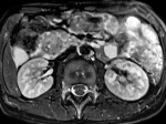
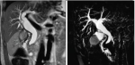
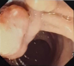
Diagnostic assessment: there was no biological inflammatory syndrome or disturbance in liver and pancreatic balance. The determination of tumor markers such as CA 19-9, ACE, and alpha-fetoprotein was negative. An ultrasound was performed showing dilatation of the bile ducts upstream of a mass in the pancreaticoduodenal region. A complementary CT revealed hypodense pancreatic cephalic mass with low enhancement after injection, measuring 21x26x5.1 mm and well-defined, resulting in significant dilatation of the main bile duct (measured at 18 mm) and the intrahepatic bile ducts ( Figure 1). Given the terrain and appearance of the CT scan, malformation pathology was suspected. MRI shows a hypointense mass on T1, hyperintense on T2, and moderate enhancement after gadolinium injection ( Figure 2). A lateral duodenoscopy was ordered, showing normal major and accessory papillae with the presence of a deep, irregularly contoured ulcerated process with an elevated bulge of 22 mm in diameter located just below the major papilla and on the medial border of D2 ( Figure 3). A duodenal biopsy and a biopsy of the detected mass were performed.
Diagnosis: duodenal biopsies and immunohistochemical tests are in favor of a moderately differentiated adenocarcinoma of the second duodenum infiltrating the head of the pancreas.
Therapeutic intervention: in front of this duodenal tumor that invades the pancreas, the surgical indication was presented at multidisciplinary consultation meetings. Peroperative investigation showed a poorly limited mass of the duodenum, measuring about 03 cm, that infiltrated the pancreatic head. A cephalic duodenopancreatectomy was performed under general anesthesia, removing the entire tumor in one piece ( Figure 4). Due to the complexity of the surgical procedure, the patient was closely monitored postoperatively in an intensive care unit for the first 24 hours, after which she was transferred to our surgical ward.
Follow-up and outcome of interventions: the postoperative course was simple with the correct glycemic cycles. Refeeding on day 2 after the operation was well tolerated. The child was discharged on day 5 after the operation. Pathological examination of the operative specimen concluded a 4 cm well-differentiated duodenum adenocarcinoma infiltrating the ampulla of Vater, extending deep into the underlying pancreas sparing the peripancreatic fat with no signs of peri-nervous engorgement or vascular invasion. All surgical borders are intact. The lymph node survey showed no lymph node metastasis. The tumor was classified as T4N0Mx. The postoperative ultrasound was normal and the current follow-up is 3 months without complications.
Patient perspective: the patient and her parents expressed their gratitude and satisfaction with the care she received. They reported feeling reassured throughout the process, from diagnosis to surgery, and appreciated the clear explanations provided by the medical team. The attentive postoperative monitoring and support helped them feel safe and confident in her recovery following the pancreaticoduodenectomy for duodenal adenocarcinoma. They emphasized their overall satisfaction with the management and the outcome of the treatment, highlighting the professionalism and dedication of the healthcare team.
Informed consent: the patient is a minor and therefore did not provide consent herself. Her parents have read and understood the information provided in the consent statement. They have voluntarily granted permission for the publication of their daughter’s medical case report as described. They acknowledge that all personal identifiers will remain confidential and that they retain the right to withdraw their consent at any time prior to publication.
Malignant tumors of the gastrointestinal tract are rare in children; duodenal adenocarcinomas are even rarer and represent less than 1% [1, 2]. In our patient, the diagnosis was particularly unexpected given her young age (11 years old) and the complete absence of personal or family history suggestive of genetic predisposing syndromes such as Lynch syndrome or familial adenomatous polyposis (FAP). The diagnosis is often made at an advanced stage due to the nonspecificity of the clinical symptoms [3]. The clinical signs are diverse: abdominal pain, melena, weight loss, vomiting, jaundice, and in most cases, the discovery of the tumor is fortuitous [4-6]. Unlike typical adult presentations where weight loss and jaundice predominate, our patient presented with isolated right hypochondrium pain and postprandial vomiting without weight loss or altered general status, reflecting the diagnostic difficulty in pediatric cases. Esogastroduodenoscopy with biopsy is a key exam for the definitive diagnosis [2]. However, radiological findings are essential to define the grade and therapeutic strategy [3]. In our case, cross-sectional imaging initially pointed toward a benign pancreaticoduodenal malformation-a common diagnostic pitfall in children-before duodenoscopy revealed an ulcerated lesion below the major papilla.
Curative management is surgical. Cephalic duodenopancreatectomy with monobloc tumor removal remains the gold standard for D1 and D2 tumors. Although these tumors are not very sensitive to chemotherapy, the role of neoadjuvant chemotherapy is being evaluated for locally advanced tumors [2]. Despite the technical challenge and high complexity of a duodenopancreatectomy in an 11-year-old child, complete monobloc resection was successfully achieved without postoperative complications, allowing early discharge on postoperative day 5. The 5-year survival rate is estimated to be 60% for curatively resected adenocarcinomas [2, 4]. The diagnosis is often made at an advanced stage. Early diagnosis and curative surgery are often correlated with longer overall survival [2]. Our case highlights that despite a locally advanced T4 stage infiltrating the pancreas, early radical surgical management can achieve complete pathological clearance, yielding a favorable short-term outcome at a 3-month follow-up.
Primary duodenal adenocarcinoma in an 11-year-old girl was successfully managed with complete R0 cephalic duodenopancreatectomy despite initial cross-sectional imaging misidentifying the mass as a congenital malformation. This case demonstrates that primary duodenal malignancies, although exceptional in pediatric patients, must be considered in the differential diagnosis of persistent upper gastrointestinal pain and vomiting. Maintaining a high index of clinical suspicion when faced with atypical pancreaticoduodenal masses is critical to avoiding diagnostic delays, as early radical surgical resection with clear margins remains the cornerstone for achieving long-term disease-free survival.
The authors declare no competing interests.
Patient management: Wael Boujelbène, Amina Chaka and Islem Bradai. Data collection: Wael Boujelbène, Amina Chaka, Amin Zouari and Amine Chaabouni. Manuscript drafting: Wael Boujelbène, Amina Chaka, Nizar Kardoun and Salma Ketata. Manuscript revision: Wael Boujelbène, Amina Chaka and Salah Boujelbène. All authors read and approved the final manuscript.
Figure 1: the abdominal contrast-enhanced computed tomography scan reveals a well-limited, hypo-intense pancreatic cephalic mass (21x26x5.1) with low post-injection enhancement;this lesion is responsible for a significant dilatation of both the intrahepatic and extrahepatic bile ducts
Figure 2: the magnetic resonance imaging demonstrates a moderately enhancing, T2-weighted hyperintense mass located at the pancreaticobiliary junction, associated with a characteristic double-duct dilatation
Figure 3: the lateral duodenoscopy shows a deep, irregular, and ulcerated mucosal lesion located beneath the major papilla in the second part of the duodenum
Figure 4: surgical specimen obtained after a successful cephalic duodenopancreatectomy (Whipple procedure), demonstrating the complete, en bloc resection of the tumor
- Tsuzuki Y, Ooshiro K, Tatekawa Y, Tamashiro R, Yagi T, Higa T. An innovative surgical approach for solid pseudopapillary neoplasm with duodenal invasion in a pediatric patient: a case report. Surg Case Rep. 2024 Oct 24;10(1):243. PubMed | Google Scholar
- Mohamed Z, Habib B, Rabia B, Youssef H, Riath B, Youssef G et al. Duodenal adenocarcinoma in a 10-year-old boy. Afr J Paediatr Surg. 2014 Jan-Mar;11(1):79-81. PubMed | Google Scholar
- Cloyd JM, George E, Visser BC. Duodenal adenocarcinoma: Advances in diagnosis and surgical management. World J Gastrointest Surg. 2016 Mar 27;8(3):212-21. PubMed | Google Scholar
- Fu H, Li Y, Bai G, Yin R, Yin C, Shi W et al. Persistent cholestasis resulting from duodenal papillary carcinoma in an adolescent male: A case report. Medicine (Baltimore). 2019 May;98(22):e15708. PubMed | Google Scholar
- Hadj Kacem H, Dafiri R. Causes tumorales d'ictére cholestatique chez l'enfant. Feuillets de radiologie. 2010 Jun 1;50(3):107-14. Google Scholar
- Alomar K, Orabi A, Qatleesh S, Saleh IEA, Hamdan O, Ahmad F. A rare case of Burkitt’s lymphoma of the duodenal bulb in a 9 year-old child - A case report and review of the literature. Int J Surg Case Rep. 2023 Aug:109:108525. PubMed | Google Scholar
Search
This article authors
On Pubmed
On Google Scholar
Citation [Download]
Navigate this article
Similar articles in
Key words
Tables and figures

Article metrics
Recently from the PAMJ







Authors´ services
