
Case of a 30-year-female with systemic lupus erythematosus: a rare clinical image
Switi Jawade, Pratibha Wankhede
Corresponding author: Switi Jawade, Nursing Tutor Florence Nightingale Training College of Nursing, Datta Meghe Institute of Medical Science (DU) Sawangi, Wardha, Maharashtra, India 
Received: 29 Jun 2022 - Accepted: 01 Aug 2022 - Published: 18 Aug 2022
Domain: Emergency medicine
Keywords: Systemic lupus erythematosus, connective tissue disorder, autoimmune disease
©Switi Jawade et al. Pan African Medical Journal (ISSN: 1937-8688). This is an Open Access article distributed under the terms of the Creative Commons Attribution International 4.0 License (https://creativecommons.org/licenses/by/4.0/), which permits unrestricted use, distribution, and reproduction in any medium, provided the original work is properly cited.
Cite this article: Switi Jawade et al. Case of a 30-year-female with systemic lupus erythematosus: a rare clinical image. Pan African Medical Journal. 2022;42:290. [doi: 10.11604/pamj.2022.42.290.36118]
Available online at: https://www.panafrican-med-journal.com//content/article/42/290/full
Images in clinical medicine 

Case of a 30-year-female with systemic lupus erythematosus: a rare clinical image
Case of a 30-year-female with systemic lupus erythematosus: a rare clinical image
Switi Jawade1,&, Pratibha Wankhede2
&Corresponding author

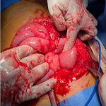
Systemic lupus erythematosus is a chronic and autoimmune disease that causes inflammation in connective tissue such as cartilage and the lining of blood vessels in which the immune system attacks its tissues. The aetiology factors are unknown but are linked to environmental, genetic, and hormonal factors; they can affect the joints, skin, brain, lungs, kidneys and blood vessels. We here report the case of a 30-year-female patient, who came to the dermatology ward with the complaints of flat red rash across the cheek and bridge of the nose, fluid-filled lesion all over the body, pain and itching, burning associated with the lesion, pedal oedema, oral ulcer, she was apparently alright four days back when she developed fluid-filled lesion which was sudden in onset and gradually progressive, the lesion occurred after ingestion of drug for fever. Initially, blood, electrocardiogram (ECG), chest X-ray investigation done, ultrasound sonography test (USG) done, which was suggestive of mild hepatosplenomegaly with the dilated portal vein and minimal free fluid in the pouch of Douglas, raised cortical echotexture of kidneys, skin biopsy done, and it shows atrophied epidermis, histopathological features suggestive of erythema multiform. The patient was referred to the dermatology and medicine department for further management.
Figure 1: systemic lupus erythematosus
Search
This article authors
On Pubmed
On Google Scholar
Citation [Download]
Navigate this article
Similar articles in
Key words
Tables and figures

Article metrics
Recently from the PAMJ


Authors´ services
