
Complete cervical agenesis: successful surgical treatment: one case report
Soumaya Kraiem, Olfa Zoukar, Asma Hnayin, Ahmed Zouari, Raja Faleh, Anis Haddad
Corresponding author: Soumaya Kraiem, Department of Gynecology Obstetric, Maternity and Neonatology Center of Monastir, University of Monastir, Monastir, Tunisia 
Received: 18 Jun 2020 - Accepted: 20 Jun 2020 - Published: 23 Jul 2020
Domain: Gynecology
Keywords: Amenorrhea, congenital cervical agenesis, reconstructive surgical procedure
©Soumaya Kraiem et al. Pan African Medical Journal (ISSN: 1937-8688). This is an Open Access article distributed under the terms of the Creative Commons Attribution International 4.0 License (https://creativecommons.org/licenses/by/4.0/), which permits unrestricted use, distribution, and reproduction in any medium, provided the original work is properly cited.
Cite this article: Soumaya Kraiem et al. Complete cervical agenesis: successful surgical treatment: one case report. Pan African Medical Journal. 2020;36:211. [doi: 10.11604/pamj.2020.36.211.24408]
Available online at: https://www.panafrican-med-journal.com//content/article/36/211/full
Case report 

Complete cervical agenesis: successful surgical treatment: one case report
Complete cervical agenesis: successful surgical treatment: one case report
Soumaya Kraiem1,&, Olfa Zoukar1, Asma Hnayin1, Ahmed Zouari1, Raja Faleh1, Anis Haddad1
&Corresponding author
Cervical agenesis is a rare congenital pathology linked to an abnormality in the development of the Mullerian system, the mechanism of this anomaly is unknown. We reported a case of complete cervical agenesis in a 17-year-old girl who underwent a successfully utero-vaginal anastomosis.

Cervical agenesis is a rare Mullerian anomaly with the incidence of 1 in 80,000 to 1,00,000 live births [1]. This is a situation in which the cervix is absent and the isthmus (the lower segment of the uterus) narrows and closes, ending in the peritoneal sleeve above and away from the vaginal apex. Patients affected by this rare abnormality have a functional uterus, but without their cervix they get primary amenorrhea with cyclic pelvic pain du to hematometra. The challenge for the clinician is to restore normal periods and potentially preserve fertility.

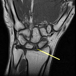
A 17-year-old virgin girl referred to our tertiary center with a story of primary amenorrhea and severe cyclic abdominal pain occuring over a period of three years. General examination results were normal and secondary sexual characteristics were well developed. The hymen was intact and had normal perforations. Ultrasound was suggestive of hematometra with right endometrioma of 7 x 7cm. Laparoscopy was proposed and showed a low abundance of brownish blood suggesting tubal reflux, the uterus and appendix were normal. A cystectomy was done. Magnetic resonance imaging (MRI) showed a normal endometrial cavity and the absence of cervical canal; the vagina was normal (Figure 1). Informed consent from the patient's parents was obtained after verbal counseling and an explanation of the potential risks of the surgery. A laparotomy was performed under general anesthesis. Intra-operatively, uterus was enlarged with normal bilatéral fallopian tubes, ovaries were normal. The proximal end of vagina was exposed after dissection of the visceral peritneum between uterus and the bladder. Midline incision in the isthmus was done to reach the caudal portion of endometrial cavity. A utero-vaginal anastomosis was successfully performed (Figure 2). The patient´s postoperative course was without problems. Menses were observed the first month after the operation. Subsequently, our patient continued to have regular periods while we followed her for the next six months. The pelvic ultrasound examination was normal.
The incidence of cervical agenesis is 0.01% in general population [1]. It represents about 3% of all uterine anomalies [2]. It is classified as type IB in the American fertility society classification of mullerian anomalies [3]. According to the new European Society of Human Reproduction and Embryology (ESHRE) and European Society for Gynaecological Endoscopy (ESGE) classification systems cervical agenesis is classified as U0C4V0 [4]. It is rarely associated with the presence of normal vagina and functioning uterus and if associated with functioning uterus, hematometra will occur. Approximately, 4.8% of women with cervical agenesis have a functioning uterus [5]. Various imaging tools have been used to assess anomalies of the Mullerian canals. Currently, magnetic resonance imaging (MRI) is the best choice for the definitive diagnosis and classification of these anomalies [6]. The conservative surgical approach of these patients involves a uterovaginal anastomosis. In fact, these patients generally preserve their fertility and need a procedure that supports cyclical rules and does not allow restenosis [7]. Unfortunately, hysterectomy might be necessary when the conservative treatment fails [8]. Cervicoplasty with mucosal lining permits the creation of a patent cervical canal, even in the reputedly unfavorable forms of congenital cervical agenesis [9].
Cervical agenesis is an anomaly that must be considered in front of any primary amenorrhea despite its rarity and this to start an adequate management because the impact on the reproductive potential of a woman can be significant.
The authors declare no competing interests.
OZ and SK conducted the literature searches; AZ and AH wrote the initial draft; AH revised the draft manuscript. All the authors have read and agreed to the final manuscript.
Figure 1: magnetic resonance imaging scan showing the vagina (in red), the isthmus (in green) and the absence of cervix (in yellow)
Figure 2: surgical view: (A) creation of a utero vaginal canal; (B) anastomosis site
- Suganuma N, Furuhashi M, Moriwaki T, Tsukahara S, Ando T, Ishihara Y. Management of missed abortion in a patient with congenital cervical atresia. Fertil Steril. 2002;77(5):1071-1073. PubMed | Google Scholar
- Grimbizis GF, Camus M, Tarlatzis BC, Bontis JN, Devroey P. Clinical implications of uterine malformations and hysteroscopic treatment results. Hum Reprod Update. 2001;7(2):161-74. PubMed | Google Scholar
- The American Fertility Society. Classification of adnexal adhesion, distal tubal occlusion secondary to tubal ligation, tubal pregnancies, Mullerian anomalies and intrauterine dhesions. Fertil Steril. 1988;49(6):944-55. PubMed | Google Scholar
- Grigoris FG, Gordts S, Di Spiezio Sardo A, Brucker S, De Angelis C, Gergolet M et al. The ESHRE/ESGE consensus on the classification of female genital tract congenital anomalies. Gynecol Surg. 2013;10(3):199-212. PubMed | Google Scholar
- Olpin JD, Heibrun M. Imaging of Mullerian duct anomalies. Clin Obstet Gynecol. 2009;52(1):40-56. PubMed | Google Scholar
- EL-Gharib MN, Awara AM. Abdominal surgical management of partial cervical agenesis in a virgin. Medical Practice and Review. 2012;3(1):5-8. Google Scholar
- Zafarani F, Ahmadi F, Shahrzad Gh. Hysterosalpingography in the assessment of congenital cervical anomalies. Int J Fertil Steril. 2017;11(2):71-78. PubMed | Google Scholar
- Achour R, Basly M, Ben-Aissa I, Ferjaoui A, Neji K. Successful pregnancy by IVF in a patient with congenital cervical atresia. Asian Pacific J Reprod. 2015;4(3):249-50. Google Scholar
- Bugmann P, Amaudruz M, Hanquinet S, La Scala G, Birraux G, Le Coultre C. Uterocervicoplasty with a bladder mucosa layer for the treatment of complete cervical agenesis Author links open overlay panel. Fertility and Sterility. April 2002;77(4):831-835. PubMed | Google Scholar
Search
This article authors
On Pubmed
On Google Scholar
Citation [Download]
Navigate this article
Similar articles in
Key words
Tables and figures


Article metrics
Recently from the PAMJ


Authors´ services
