
Exceptional intrarenal pseudomembranous
Souhail Regragui, Gabriel Stoica
Corresponding author: Souhail Regragui, Urology Departement, CHIC Alencon Mamers, Alencon, France 
Received: 27 Feb 2019 - Accepted: 11 Apr 2019 - Published: 29 Apr 2019
Domain: Urology
Keywords: Pseudomembranous, renal, pyelotomy
©Souhail Regragui et al. Pan African Medical Journal (ISSN: 1937-8688). This is an Open Access article distributed under the terms of the Creative Commons Attribution International 4.0 License (https://creativecommons.org/licenses/by/4.0/), which permits unrestricted use, distribution, and reproduction in any medium, provided the original work is properly cited.
Cite this article: Souhail Regragui et al. Exceptional intrarenal pseudomembranous. Pan African Medical Journal. 2019;32:214. [doi: 10.11604/pamj.2019.32.214.18385]
Available online at: https://www.panafrican-med-journal.com//content/article/32/214/full
Original article 

Exceptional intrarenal pseudomembranous
Exceptional intrarenal pseudomembranous
Souhail Regragui1,2,&, Gabriel Stoica1
1Urology Departement, CHIC Alencon Mamers, Alencon, France, 2Urology B Departement, CHU Ibn Sina, Rabat, Morocco
&Corresponding author
Souhail Regragui, Urology Departement, CHIC Alencon Mamers, Alencon, France

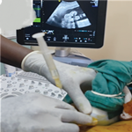
We report the case of a 51-year-old patient, known as hypertensive and type 2 diabetic, admitted to emergency for acute pyelonephritis. She suffered from low back pain in a feverish context. A Uro-scanner showed the presence of a 12mm pyelic renal calculus and a calycal calculus of 6mm diameter responsible for a moderate dilation of left pyelocalictic cavities. First, we performed drainage with a double J probe. Then, in a second step, the left ureteroscopy allowed partial laser fragmentation. The presence of a suspect soft magma prompted us to stop the procedure. After performing a hydatid serology that has returned negative, a laparoscopic left pyelotomy allowed the progressive externalization of the suspect magma. It presented with a greenish, fibrinous, semi-solid shell completely molding the pyelon and the pyelocalicielles cavities. The introduction of the flexible cystoscope through a trocar at the end of the procedure allowed us to check the complete cleaning of the excretory cavities. The anatomopathological study is back in favor of a weakly eosinophilic acellular material with some polyhedral crystals and some inflammatory elements. There were no signs of malignancy. The patient did not present any postoperative complication and did not present a recurrence.
Figure 1: A) extraction of the pseudomembranous from the kidney; B) final aspect of the pseudomembranous
Search
This article authors
On Pubmed
On Google Scholar
Citation [Download]
Navigate this article
Similar articles in
Key words
Tables and figures

Article metrics
PlumX Metrics
Exceptional intrarenal pseudomembranousRecently from the PAMJ


Authors´ services
