
eISSN: 1937-8688

Pulmonary arteriovenous malformation and follow-up imaging

Pahnwat Tonya Taweesedt, Salim Surani

PAMJ. 2020; 37:294. Published 02 Dec 2020 | DOI: 10.11604/pamj.2020.37.294.27100
Pulmonary arteriovenous malformation and follow-up imaging
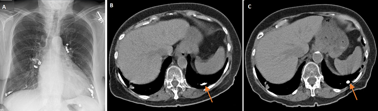
An 85-year-old Caucasian female with past medical history of hypertension, hyperlipidemia, polymyalgia rheumatica, coronary artery disease, Osler-Weber-Rendu syndrome (diagnosed 18 years ago), intermittent epistaxis and pulmonary arteriovenous malformation (AVM) was referred to the clinic for abnormal chest imaging. She underwent multiple bilateral AVM embolization 17 years ago. She had minimal shortness of breath without epistaxis, hemoptysis, hematemesis or hematochezia. One month ago, her chest X-ray revealed multiple embolization coils bilaterally (A). Computed tomography (CT) chest scan without contrast showed multiple embolization coils, multiple tortuous vascular malformations in both lungs with enlargement of left lower lobe AVM (B). Magnetic resonance imaging (MRI) brain 17 years ago did not show brain vascular malformation. The diagnosis of progressive Osler-Weber-Rendu or hereditary hemorrhagic telangiectasia (HHT) was made. She was referred to HHT center and underwent coiling. After that, she has been clinically stable and a repeat CT chest on the follow-up visit showed smaller left lower lobe AVM (B), so conservative management was planned.

Corresponding Author

Pahnwat Tonya Taweesedt, Department of Pulmonary Medicine, Corpus Christi Medical Center, Texas, United States of America (Pahnwatt@gmail.com)

Article Formats

Articles published in Pan African Medical Journal are Open Access and distributed under the terms of the Creative Commons Attribution 4.0 International (CC BY 4.0).

Open Access Logo

The Pan African Medical Journal (ISSN: 1937-8688) is a product of the PAMJ. The contents of this journal is intended exclusively for professionals in the medical, paramedical and public health and other health sectors.