
Assessment of ureteral dimensions on contrast-enhanced multidetector computed tomography of the urinary tract
Ngo Xuan Khoa, Vo Tien Huy, Ngo Thuy Anh Ngoc, Tran Quoc Hoa, Nguyen Ngoc Anh
Corresponding author: Vo Tien Huy, VNU University of Medicine and Pharmacy, Vietnam National University, Hanoi, Vietnam 
Received: 17 Feb 2026 - Accepted: 26 Jun 2026 - Published: 07 Jul 2026
Domain: Radiology
Keywords: Ureters, computed tomography, dimensions
Funding: This work received no specific grant from any funding agency in the public, commercial, or non-profit sectors.
This article is published as part of the supplement Innovations and Challenges in Global Health: A Multidisciplinary Perspective, commissioned by Young Researchers and Elite Club.
©Ngo Xuan Khoa et al. Pan African Medical Journal (ISSN: 1937-8688). This is an Open Access article distributed under the terms of the Creative Commons Attribution International 4.0 License (https://creativecommons.org/licenses/by/4.0/), which permits unrestricted use, distribution, and reproduction in any medium, provided the original work is properly cited.
Cite this article: Ngo Xuan Khoa et al. Assessment of ureteral dimensions on contrast-enhanced multidetector computed tomography of the urinary tract. Pan African Medical Journal. 2026;54(1):15. [doi: 10.11604/pamj.supp.2026.54.1.51675]
Available online at: https://www.panafrican-med-journal.com//content/series/54/1/15/full
Research 

Assessment of ureteral dimensions on contrast-enhanced multidetector computed tomography of the urinary tract
Assessment of ureteral dimensions on contrast-enhanced multidetector computed tomography of the urinary tract
Ngo Xuan Khoa1,2, Vo Tien Huy3,4,&, Ngo Thuy Anh Ngoc5, Tran Quoc Hoa1, Nguyen Ngoc Anh1
&Corresponding author
Introduction: the objective to determine the anatomical dimensions of the ureter on contrast-enhanced multidetector computed tomography (MDCT) of the urinary tract and to assess their relationship with selected anthropometric parameters.
Methods: this was a cross-sectional descriptive study conducted on 102 patients (61 males, 41 females) without a history of urinary tract trauma, surgery, or intervention, and with at least one anatomically normal ureter, as confirmed by radiologists. All participants underwent contrast-enhanced MDCT urography between January 2024 and March 2025 at Hanoi Medical University Hospital.
Results: the mean ureteral length was 22.56 ± 2.55cm on the right and 23.28 ± 3.82cm on the left, with no statistically significant differences between sides or sexes. The average ureteral diameter at natural constriction points ranged from 2.8 to 3.39mm. No significant correlations were found between ureteral dimensions and height, weight, or body mass index (p > 0.05).
Conclusion: the findings provide valuable reference data for radiologic evaluation and the selection of appropriate ureteral stent lengths in clinical practice.

The evaluation of ureteral diameter plays an essential role in diagnosing and managing various urinary tract conditions, including ureterolithiasis, congenital anomalies, and upper tract urothelial carcinoma (UTUC) [1,2]. Among imaging modalities, multidetector computed tomography (MDCT) has emerged as a preferred method due to its high spatial resolution, cross-sectional capability, and superior diagnostic performance [2,3]. MDCT enables detailed visualisation of both the anatomy and pathology of the urinary system, making it a valuable tool in clinical decision-making. It offers the ability to assess ureteral calibre, detect filling defects, and evaluate secondary signs of obstruction, which are crucial for prompt and accurate treatment planning [3,4].
Understanding normal ureteral dimensions is critical for identifying pathological changes. Zelenko et al. reported that the mean ureteral diameter on the asymptomatic side is approximately 1.8mm, with notable inter-individual variability [5]. This knowledge provides a reference point for detecting ureteral dilatation-an important indirect sign of obstruction in cases such as stone disease. Furthermore, evaluating the ureter's size helps determine the need for intervention, monitor treatment outcomes, and assess anatomical variations that may influence surgical or endoscopic approaches [1]. Accurate reference values are also important for selecting appropriate ureteral stent lengths and avoiding unnecessary overestimation of ureteral dilatation during radiologic interpretation.
MDCT urography has been widely recognised for its accuracy in diagnosing upper urinary tract conditions. According to Potenta et al., MDCT provides comprehensive information on the urinary tract's morphology, detecting pathologies such as strictures, filling defects, or dilated segments [1]. In the context of UTUC, Janisch et al. highlighted the high diagnostic performance of MDCT, reporting pooled sensitivity and specificity of 92% and 95%, respectively [6]. Moreover, MDCT is valuable in assessing uroepithelial malignancies and urinary calculi, with its ability to evaluate stone size, location, and composition. These characteristics are essential in predicting spontaneous passage, particularly for small stones [3,7], and advanced techniques like dual-energy CT (DECT) and virtual non-contrast CT (vNCT) further enhance the evaluation [4,8].
Despite its advantages, MDCT interpretation must consider potential limitations. For example, Shaish et al. noted that while split-bolus CT protocols offer high sensitivity, their positive predictive value can be low, requiring cautious analysis [9]. Moreover, concerns related to cost, radiation exposure, and disease prevalence must be balanced against diagnostic needs, especially in screening for conditions like UTUC. Although normative ureteral measurements have been reported in several populations, substantial variability exists across studies because of differences in ethnicity, imaging protocols, measurement techniques, and anatomical landmarks. To date, comprehensive MDCT-based reference data for normal ureteral dimensions in the Vietnamese population remain limited. Establishing population-specific reference values may improve the interpretation of urinary tract imaging and support more appropriate preoperative planning for ureteral interventions. Therefore, this study aimed to determine normal ureteral dimensions on contrast-enhanced MDCT and evaluate their associations with sex and anthropometric characteristics in a Vietnamese population.

Study design: this descriptive cross-sectional study investigated normal ureteral dimensions and their associations with anthropometric characteristics using multidetector computed tomography (MDCT) imaging in a Vietnamese population.
Setting: the study was conducted at Hanoi Medical University Hospital, a tertiary academic medical centre in Vietnam, over 15 months from January 2024 to March 2025. All radiologic examinations and clinical data were obtained from routine diagnostic services within the hospital.
Participants: the study population consisted of Vietnamese patients who underwent contrast-enhanced MDCT imaging of the urinary system during the study period. Patients were eligible if at least one ureter was confirmed to be anatomically and radiologically normal by expert radiologic assessment, with complete medical records and MDCT images of sufficient quality to clearly visualise the ureter and adjacent anatomical structures. Patients with a history of urinary tract trauma, prior urologic surgery or intervention, or suboptimal imaging due to artefacts or noise were excluded. Using convenience sampling, 102 patients meeting all eligibility criteria were included.
Variables: primary variables were ureteral length and ureteral diameter measured at predefined anatomical levels. Covariates included demographic and anthropometric characteristics (age, sex, height, and weight). The study also examined correlations between ureteral dimensions and anthropometric indices.
Data sources and measurement: demographic and anthropometric data were retrieved from the hospital information system. Radiologic data were obtained from the picture archiving and communication system and included contrast-enhanced MDCT scans acquired using 16-slice and 128-slice scanners. Ureters were evaluated on delayed-phase images with multiplanar reconstruction and maximum intensity projection techniques to optimise visualisation. Ureteral length was measured along the curved ureteral course in segments, and ureteral diameter was measured on cross-sectional images using electronic callipers at predetermined levels.
Bias: selection bias was minimised by applying predefined inclusion and exclusion criteria and including all eligible examinations during the study period. Measurement bias was reduced through standardised MDCT protocols, use of advanced reconstruction techniques, and expert radiologic assessment. However, potential bias related to convenience sampling and single-centre design may limit generalizability.
Study size: the study sample comprised all eligible patients undergoing MDCT urography at Hanoi Medical University Hospital during the study period, resulting in a final sample of 102 participants.
Quantitative variables: continuous variables, including ureteral length, ureteral diameter, age, height, and weight, were summarised as means with standard deviations. Categorical variables, such as sex, were expressed as frequencies and percentages. Anthropometric variables were analysed as continuous measures in correlation analyses.
Statistical methods: data were entered and analysed using SPSS version 20.0. Descriptive statistics summarised demographic characteristics and ureteral measurements. Associations between ureteral dimensions and anthropometric indices were evaluated using appropriate correlation tests. Statistical significance was defined as a two-sided p-value <0.05.
Ethical consideration statement: the study protocol was approved by the Institutional Ethics Committee of Hanoi Medical University (Decision No. 3658/QÐ-ÐHYHN). All procedures involving human participants were conducted in accordance with institutional ethical standards and the 2013 Declaration of Helsinki, and all data were analysed in anonymised form.
Demographic characteristics: among 102 patients, the average age of participants was 49.43 ± 15.41 years, with a relatively even distribution between those aged 20-50 years and those over 50 years. Males made up a higher proportion of the study sample (59.8%). The average weight and height were 59.02 kg and 1.63 m, respectively, while the average BMI was 22.10 kg/m², corresponding to the normal BMI range (Table 1).
Descriptive statistics: Table 2 presents the measured lengths of the abdominal and pelvic segments of the ureter by side and gender. The mean length of the abdominal ureter on the right side was 12.63 ± 1.46cm in males and 11.34 ± 1.27cm in females, with a combined average of 12.51 ± 1.61cm; on the left side, it was 12.65 ± 1.69cm in males and 12.42 ± 1.54cm in females, with a combined mean of 12.56 ± 1.83cm. For the pelvic segment, the right ureter measured 10.56 ± 1.23cm in males and 10.13 ± 1.57cm in females (combined: 10.15 ± 1.62cm), while the left side measured 11.30 ± 1.55cm in males and 10.35 ± 1.39cm in females (combined: 10.73 ± 1.74cm). The p-values indicate no statistically significant differences between genders or between left and right sides for either segment.
Table 3 presents the total length of the right and left ureters measured in the study participants. The average length of the right ureter was 22.90 ± 2.64cm in males and 22.46 ± 2.19cm in females, with a combined mean of 22.56 ± 2.55cm. For the left ureter, the average length was 23.72 ± 3.22cm in males and 23.00 ± 2.71cm in females, with a combined mean of 23.28 ± 3.82cm. There were no statistically significant differences in ureteral length between genders or between the left and right sides.
Table 4 shows the ureteral diameter measured at three natural constriction points: the ureteropelvic junction (UPJ), the point where the ureter crosses the iliac vessels, and the ureterovesical junction (UVJ). The average diameter at the UPJ ranged from 3.24mm to 3.55mm, with no statistically significant differences between sexes or between left and right sides. At the iliac vessel crossing, the mean diameter varied from 3.07mm to 3.22mm, and at the UVJ, it ranged from 2.74mm to 2.94mm. In all locations, the p-values indicated no significant gender-based or side-specific differences.
Table 5 shows that no statistically significant correlation was observed between body indices (height, weight, and BMI) and ureteral length or diameter, as all p-values were greater than 0.05.
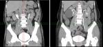
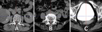
MDCT images: Figure 1 presents coronal MDCT images of two patients illustrating the extremes in ureteral length within the study sample. The image on the left shows the patient with the shortest measured ureteral length, while the image on the right shows the patient with the longest ureteral length. Both ureters are visualised from the renal pelvis to the bladder, with measurement lines indicating the course and curvature of the ureters. This comparison highlights the anatomical variability in ureteral length among individuals. Figure 2 illustrates the measurement of ureteral diameter at different sites of physiological narrowing. In all images, the ureter is clearly visualised, and electronic callipers are used to determine the transverse diameter. These anatomical landmarks are critical for assessing normal ureteral calibre and identifying potential sites of obstruction.
This study investigated the anatomical dimensions of the ureter, including total length, segmental length, and diameter at natural constriction points, in a Vietnamese population using multidetector computed tomography (MDCT). The findings demonstrated consistent measurements across gender and laterality, with no statistically significant differences between males and females or between right and left sides in any of the evaluated parameters. These findings provide normative MDCT-based reference values for the Vietnamese population and contribute to the limited anatomical imaging data currently available for this population.
According to the study findings, the mean length of the ureter in the abdominal and pelvic segments was approximately 12.5cm and 10.15-11.30cm, respectively. Compared with classical anatomical descriptions, which report ureteral lengths ranging from 12.5 to 14cm, the abdominal segment length in our study was consistent, whereas the pelvic segment was 1-2.5cm shorter. The total ureteral lengths for the right and left sides in our study were 22.56 ± 2.55cm and 23.28 ± 3.82cm, respectively. These values are comparable to the results of Shah et al. [10], who reported a mean ureteral length of 23.46cm measured using cystoscopy, and slightly lower than the 25cm reported by Jung et al. in Korea [11], who measured ureteral length using MPR-reconstructed CT images along the ureter's curved course. These modest differences are likely attributable not only to differences in measurement techniques but also to population characteristics and imaging protocols. Our measurements were performed manually by segmenting the ureter along its curvature and summing the individual lengths, which may account for methodological differences compared to Jung's study. Additionally, our findings revealed no statistically significant differences in ureteral length between the left and right sides or between male and female participants, consistent with the findings of Jung et al. [11]. Although the left ureter was numerically longer than the right, this difference was not statistically significant and is anatomically plausible because the left kidney is typically positioned slightly higher than the right, resulting in a longer ureteral course.
In this study, we also measured ureteral diameter at three natural constriction points: the UPJ, the point where the ureter crosses the iliac vessels, and the UVJ. The average diameters at these locations ranged from approximately 2.8mm to 3.3mm, with no significant differences between sides or between sexes. These values were lower than those reported in the study by Itanyi et al. in Nigeria [12], where average diameters ranged from 4.3 to 4.4mm, measured on contrast-enhanced abdominal CT scans, and also lower than those found by Wong et al. in Malaysia [13], who reported values between 4.2 and 4.5mm using intravenous urography. This discrepancy is likely due to the fact that our study focused on the diameter at natural constriction points, while the Itanyi et al. study measured 1-2cm below the UPJ, and Wong's measurements were taken within 6cm below the UPJ. Differences in imaging protocols, patient preparation, bladder filling, ureteral peristalsis, and the timing of contrast-enhanced image acquisition may also have contributed to the variability among studies. Interestingly, our diameter results were higher than those reported by Zelenko et al. [5] on non-contrast CT, where the mean ureteral diameter was 1.8 ± 0.9mm, and 3mm was proposed as the upper limit of normal. The difference is likely attributable to imaging techniques: our study used contrast-enhanced CT in the excretory phase, during which the ureter is distended by contrast medium, while the measurements in the Zelenko et al. [5] study were obtained from non-enhanced images. Nonetheless, similar to other studies, we found no significant differences in ureteral diameter at these constriction points across sides or sexes. The absence of sex-related differences suggests that ureteral calibre is relatively independent of sex despite differences in overall body size, supporting the use of common reference values for adult males and females.
Furthermore, we investigated potential correlations between ureteral dimensions and anthropometric parameters such as height, weight, and BMI. The results showed no statistically significant correlation between these body measurements and ureteral length or diameter (r < 0.1, p > 0.05). This is in agreement with studies such as that by Shah et al. [10], which found that height could not predict ureteral length (r² = 0.017, p = 0.54), and that by Jung et al. [11], who reported only a weak correlation (r² ≈ 0.06). In contrast, some European studies have traditionally used patient height to estimate ureteral length for stent selection. However, in Asian populations, including our study and others, no strong correlation has been found between these variables. This finding suggests that external body size does not reliably reflect ureteral anatomy, likely because ureteral length and calibre are influenced primarily by individual anatomical variation rather than overall body habitus. Therefore, relying solely on patient height to determine ureteral stent length may lead to inaccuracies in Asian settings.
The findings of this study provide baseline reference values for ureteral dimensions in the Vietnamese population. This information can be used by radiologists and urologists for comparison in pathological cases, aiding in the diagnosis of obstructive uropathy, congenital abnormalities, and ureteral trauma. The reported values may also facilitate more accurate interpretation of ureteral dilatation on MDCT and assist clinicians in selecting appropriate ureteral stent lengths during preoperative planning. Furthermore, the application of MDCT imaging demonstrates its practicality and accuracy in evaluating ureteral anatomy non-invasively.
A major strength of this study is that ureteral measurements were obtained using contrast-enhanced MDCT with multiplanar reconstruction, allowing assessment of the ureter along its natural anatomical course rather than by linear approximation. In addition, both ureteral length and physiological narrowing sites were systematically evaluated bilaterally, providing comprehensive reference data for clinical practice. However, this study has several limitations. First, the sample size, although sufficient for descriptive analysis, was drawn from a single tertiary hospital using convenience sampling, which may limit generalizability to the broader population. Second, while efforts were made to exclude patients with urinary tract pathology, subclinical or undiagnosed conditions may have influenced measurements. Third, the study did not include interobserver variability in ureteral measurement, which could be addressed in future research to validate reproducibility. Lastly, the supine imaging position may not fully reflect dynamic changes in ureteral length or diameter during physiologic processes such as bladder filling. In addition, hydration status and the degree of bladder distension were not standardised before imaging, factors that may influence ureteral calibre. Future multicenter studies with larger sample sizes, standardised imaging protocols, and assessment of measurement reproducibility are warranted to validate these findings and establish robust reference standards for clinical application.
This study established reference values for ureteral length and diameter at key anatomical locations using MDCT in a Vietnamese population. The results indicate no significant differences between genders or between left and right ureters, supporting the reliability of using uniform criteria in clinical evaluation. These findings enhance the anatomical knowledge base for radiological assessment and urological procedures and underscore the value of MDCT in urinary tract imaging. Future studies with larger, multi-centre cohorts and dynamic imaging modalities are recommended to further refine these anatomical standards.
What is known about this topic
- Multidetector computed tomography (MDCT) urography provides high-resolution visualisation of ureteral anatomy and is widely used to assess ureteral calibre, detect obstruction, and guide management of urinary tract diseases;
- Previous anatomical and imaging studies have reported considerable inter-individual variability in ureteral length and diameter, with generally limited or inconsistent correlations between ureteral dimensions and patient anthropometric characteristics.
What this study adds
- This study establishes MDCT-based reference values for ureteral length and diameter at key anatomical constriction points in a Vietnamese population, demonstrating no significant differences between sides or sexes;
- The findings show no meaningful correlation between ureteral dimensions and height, weight, or body mass index, indicating that anthropometric measures are unreliable predictors of ureteral size for stent selection in this population.
The authors declare no competing interests.
Ngo Xuan Khoa: study conception and design, imaging analysis, data interpretation, and manuscript drafting. Vo Tien Huy: methodological supervision, statistical analysis oversight, and critical revision of the manuscript. Ngo Thuy Anh Ngoc: data acquisition and radiologic measurements. Tran Quoc Hoa: data collection and database management. Nguyen Ngoc Anh: literature review and manuscript editing. All authors read and approved the final version of the manuscript.
The authors thank the radiology staff of Hanoi Medical University Hospital for their assistance in imaging acquisition and data retrieval.
Table 1: demographic and anthropometric characteristics of the study sample
Table 2: length of the abdominal and pelvic segments of the ureter
Table 3: total ureteral length in study participants
Table 4: ureteral diameter at natural constriction points
Table 5: correlation between body indices and ureteral length and diameter
Figure 1: comparison of shortest and longest ureteral lengths in patients
Figure 2: ureteral diameter measurement A) at the Ureteropelvic Junction (UPJ); B) at the Point Where the Ureter Crosses the Iliac Vessels; C) at the Ureterovesical Junction (UVJ)
- Potenta SE, D'Agostino R, Sternberg KM, Tatsumi K, Perusse K. CT Urography for Evaluation of the Ureter. Radiographics. 2015 May-Jun;35(3):709-26. PubMed | Google Scholar
- Houat AP, Guimarães CTS, Takahashi MS, Rodi GP, Gasparetto TPD, Blasbalg R et al. Congenital Anomalies of the Upper Urinary Tract: A Comprehensive Review. Radiographics. 2021 Mar-Apr;41(2):462-486. doi: 10.1148/rg.2021200078. Epub 2021 Jan 29. Erratum in: Radiographics. 2021 Sep-Oct;41(5):E165. PubMed | Google Scholar
- Demehri S, Steigner ML, Sodickson AD, Houseman EA, Rybicki FJ, Silverman SG. CT-based determination of maximum ureteral stone area: a predictor of spontaneous passage. AJR Am J Roentgenol. 2012 Mar;198(3):603-8. PubMed | Google Scholar
- McCoombe K, Dobeli K, Meikle S, Llewellyn S, Kench P. Sensitivity of virtual non-contrast dual-energy CT urogram for detection of urinary calculi: a systematic review and meta-analysis. Eur Radiol. 2022 Dec;32(12):8588-8596. PubMed | Google Scholar
- Zelenko N, Coll D, Rosenfeld AT, Smith RC. Normal ureter size on unenhanced helical CT. AJR Am J Roentgenol. 2004 Apr;182(4):1039-41. PubMed | Google Scholar
- Janisch F, Shariat SF, Baltzer P, Fajkovic H, Kimura S, Iwata T et al. Diagnostic performance of multidetector computed tomographic (MDCTU) in upper tract urothelial carcinoma (UTUC): a systematic review and meta-analysis. World J Urol. 2020 May;38(5):1165-1175. PubMed | Google Scholar
- Caoili EM, Cohan RH, Korobkin M, Platt JF, Francis IR, Faerber GJ et al. Urinary tract abnormalities: initial experience with multi-detector row CT urography. Radiology. 2002 Feb;222(2):353-60. PubMed | Google Scholar
- Feng P, Li G, Liang P. The value of dual-energy computed tomography (DECT) in the diagnosis of urinary calculi: a systematic review and meta-analysis of retrospective studies. PeerJ. 2023 Oct 2;11:e16076. PubMed | Google Scholar
- Shaish H, Newhouse JH. Split-bolus CT urogram: Is less more? Abdom Radiol (NY). 2017 Aug;42(8):2119-2126. PubMed | Google Scholar
- Shah J, Kulkarni RP. Height does not predict ureteric length. Clin Radiol. 2005 Jul;60(7):812-4. PubMed | Google Scholar
- Jung SI, Park HS, Yu MH, Kim YJ, Lee H, Choi WS et al. Korean ureter length: A computed tomography-based study. Investig Clin Urol. 2020 May;61(3):291-296 PubMed | Google Scholar
- Itanyi UD, Aiyekomogbon JO, Uduma FU, Evinemi MA. Assessment of ureteric diameter using contrast-enhanced helical abdominal computed tomography. African Journal of Urology. 2020 Dec;26(1):23. Google Scholar
- Wong SL, Abdul Hamid H. Observation of ureteric diameter in negative intravenous urogram in hospital universiti kebangsaan malaysia. Malays J Med Sci. 2010 Apr;17(2):4-9. PubMed | Google Scholar
Search
This article authors
On Pubmed
On Google Scholar
Citation [Download]
Navigate this article
Similar articles in
Key words
Tables and figures

This supplement
Article metrics
Recently from the PAMJ





Authors´ services
