
Pituitary metastasis: an unexpected complication in advanced cancer
Natthaphon Wonghakaeo, Thiti Snabboon
Corresponding author: Thiti Snabboon, Department of Medicine, Faculty of Medicine, Chulalongkorn University, Bangkok, Thailand 
Received: 22 Jan 2025 - Accepted: 31 Jan 2025 - Published: 04 Feb 2025
Domain: Endocrinology, Internal medicine, Oncology
Keywords: Hypopituitarism, diabetes insipidus, breast cancer, pituitary metastasis
©Natthaphon Wonghakaeo et al. Pan African Medical Journal (ISSN: 1937-8688). This is an Open Access article distributed under the terms of the Creative Commons Attribution International 4.0 License (https://creativecommons.org/licenses/by/4.0/), which permits unrestricted use, distribution, and reproduction in any medium, provided the original work is properly cited.
Cite this article: Natthaphon Wonghakaeo et al. Pituitary metastasis: an unexpected complication in advanced cancer. Pan African Medical Journal. 2025;50:41. [doi: 10.11604/pamj.2025.50.41.46622]
Available online at: https://www.panafrican-med-journal.com//content/article/50/41/full
Images in clinical medicine 

Pituitary metastasis: an unexpected complication in advanced cancer
Pituitary metastasis: an unexpected complication in advanced cancer
Natthaphon Wonghakaeo1,2, ![]() Thiti Snabboon 2,3,&
Thiti Snabboon 2,3,&
&Corresponding author

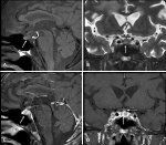
A 57-year-old man with metastatic breast cancer presented with a four-month history of polyuria, which was diagnosed as diabetes insipidus (DI). Further hormonal evaluation revealed concurrent hypothyroidism and hypocortisolism. Magnetic resonance imaging demonstrated a pituitary lesion with an absent posterior bright spot, suggesting a metastatic lesion. Unfortunately, the patient died from uncontrolled advanced disease one month later. Pituitary metastasis is rare but increasingly recognized due to advances in imaging and treatment. Breast and lung cancers are the most common primary malignancies. Clinical presentations vary from asymptomatic cases to visual field defects or pituitary dysfunction caused by compression of adjacent structures. Diabetes insipidus is the most common endocrine abnormality, with anterior hypopituitarism in 25-45% of cases. Diagnosing pituitary metastasis can be challenging, as it may be mistaken for pituitary adenoma. Key magnetic resonance imaging (MRI) findings indicative of metastasis include dumbbell-shaped lesions, stalk thickening, erosion of the sella turcica, and the absence of the posterior pituitary bright spot. The prognosis is generally poor, with a median survival of 12.9 months, and no treatment has significantly improved survival. Therefore, clinicians should consider pituitary metastasis, especially in patients with advanced malignancies presenting with DI.
Figure 1: magnetic resonance imaging findings; A) sagittal T1-weighted image showing a 1 cm hypointense lesion in the mid-posterior part of the pituitary gland (arrow), with an absent posterior bright spot (curved arrow); B) coronal T2-weighted image demonstrating a hyperintense lesion (arrow); C, D) post-gadolinium sagittal and coronal T1-weighted images revealed a heterogeneous enhancement lesion (arrow) with a normal-appearing pituitary stalk (dashed arrow); metastatic lesions were also noted in the clivus and occipital bone
Search
This article authors
On Pubmed
On Google Scholar
Citation [Download]
Navigate this article
Similar articles in
Key words
Article metrics
Recently from the PAMJ


Authors´ services
