
A rare clinical image of frontal hemorrhagic metastasis in chronic myelogenous leukaemia patient
Pradhyum Dilip Kolhe, Sharath Hullumani
Corresponding author: Sharath Hullumani, Department of Paediatric Physiotherapy, Ravi Nair Physiotherapy College, Datta Meghe Institute of Higher Education and Research (DU) Sawangi Meghe, Maharashtra, India 
Received: 07 Nov 2024 - Accepted: 06 Dec 2024 - Published: 02 Jan 2025
Domain: Neuro-oncology,Neuroradiology
Keywords: Frontal hemorrhagic metastasis, neurosurgery, oncology
©Pradhyum Dilip Kolhe et al. Pan African Medical Journal (ISSN: 1937-8688). This is an Open Access article distributed under the terms of the Creative Commons Attribution International 4.0 License (https://creativecommons.org/licenses/by/4.0/), which permits unrestricted use, distribution, and reproduction in any medium, provided the original work is properly cited.
Cite this article: Pradhyum Dilip Kolhe et al. A rare clinical image of frontal hemorrhagic metastasis in chronic myelogenous leukaemia patient. Pan African Medical Journal. 2025;50:3. [doi: 10.11604/pamj.2025.50.3.45836]
Available online at: https://www.panafrican-med-journal.com//content/article/50/3/full
Images in clinical medicine 

A rare clinical image of frontal hemorrhagic metastasis in chronic myelogenous leukaemia patient
A rare clinical image of frontal hemorrhagic metastasis in chronic myelogenous leukaemia patient
Pradhyum Dilip Kolhe1, ![]() Sharath Hullumani1,&
Sharath Hullumani1,&
&Corresponding author

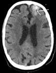
A 41-year-old male patient was brought to hospital casualty by his relatives with complaints of altered sensorium and reduced consciousness. He had a history of one episode of convulsions, involuntary movements of bilateral upper limb and lower limb. No history of bowel and bladder complaints. He had a known case of chronic myelogenous leukaemia for which he took chemotherapy ove the past 4 years. Patient was intubated in casualty. Diagnostic evaluations including a Magnetic Resonance Imaging (MRI) scan, have substantiated a diagnosis of hemorrhagic metastasis in the right frontal region. The figure revealed the presence well defined heterogeneously enhancing T2 hyperintense lesion with edema seen in right precentral gyrus measuring 13x11 mm in size metastases. A well-defined area of blooming appearing hypointense on T2 is seen in right frontal lobe known as acute hematoma metastases. Acute hematoma is seen in right basal ganglia measuring 12x8 mm in size with possible extension into bilateral, 3rd and 4th ventricle. Moderate dilatation of bilateral 3rd and 4th ventricle noted with periventricular chronic fatigue syndrome (CSF) leak. Mass effect is seen in form of midline shift of 8 mm towards left side. Patchy areas of diffusion restriction are seen in parasaggital cortex of frontoparietal lobe and body of corpus callosum likely acute infarct in bilateral anterior cerebral artery (ACA).
Figure 1: acute hematoma metastases in right frontal lobe
Search
This article authors
On Pubmed
On Google Scholar
Citation [Download]
Navigate this article
Similar articles in
Key words
Tables and figures
Article metrics
Recently from the PAMJ


Authors´ services
