
Exophthalmia revealing acute myeloid leukemia
Leila Debono, Youssef Jeddi
Corresponding author: Leila Debono, Pediatric´s Medical Emergency Department, Children´s Hospital of Rabat, Rabat, Morocco 
Received: 12 Dec 2024 - Accepted: 05 Jan 2025 - Published: 07 Jan 2025
Domain: Pediatric hematology, Pediatrics (general)
Keywords: Exophthalmia, leukemia, child
©Leila Debono et al. Pan African Medical Journal (ISSN: 1937-8688). This is an Open Access article distributed under the terms of the Creative Commons Attribution International 4.0 License (https://creativecommons.org/licenses/by/4.0/), which permits unrestricted use, distribution, and reproduction in any medium, provided the original work is properly cited.
Cite this article: Leila Debono et al. Exophthalmia revealing acute myeloid leukemia. Pan African Medical Journal. 2025;50:14. [doi: 10.11604/pamj.2025.50.14.46203]
Available online at: https://www.panafrican-med-journal.com//content/article/50/14/full
Images in clinical medicine 

Exophthalmia revealing acute myeloid leukemia
Exophthalmia revealing acute myeloid leukemia
Leila Debono1,2,&, Youssef Jeddi1,2
&Corresponding author

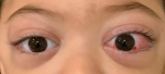
A child aged 2 years and a half, without notable history, was presented with swelling of the right eye a month previously. He consulted a general practitioner, who put him on amoxicillin with clavulanic acid for 10 days and betamethasone drops for 5 days. A week later, there was swelling in the left eye. The parents consulted an ophthalmologist who treated him for conjunctivitis and put him on tobramycin eye drops for 5 days, without improvement. The clinical examination on admission found an irritable, afebrile, eupneic child with bilateral exophthalmos greater on the left with conjunctival redness and lower right eyelid ecchymosis. The rest of the examination was normal, notably free lymph node areas and no palpable mass. A cerebral and orbital Computed Tomography (CT) scan was performed, revealing Chandler's stage I preseptal cellulitis with bilateral grade I exophthalmos; the cerebral CT scan was without abnormalities. A blood count was performed revealing bicytopenia with normocytic normochromic anemia at 6.7 g/dl with thrombocytopenia at 19,000/µl, a white blood cell level at 25,600/µl with polynuclear neutrophils at a normal level and the presence of 66% blast cells. A smear carried out showed a blood cytological appearance in favor of acute myeloid leukemia.
Figure 1: exophthalmia revealing acute myeloid leukemia
Search
This article authors
On Pubmed
On Google Scholar
Citation [Download]
Navigate this article
Similar articles in
Key words
Tables and figures
Article metrics
PlumX Metrics
Exophthalmia revealing acute myeloid leukemiaRecently from the PAMJ


Authors´ services
