
An exceptional case of a huge pseudoaneurysm caused by an epicardial electrode
Yassine Morjane, Alexandre Sebestyen
Corresponding author: Yassine Morjane, Service de Chirurgie Cardiaque, Centre Hospitalier Universitaire Grenoble-Alpes, La Tronche, France 
Received: 15 May 2023 - Accepted: 23 Aug 2023 - Published: 14 Sep 2023
Domain: Cardiovascular surgery
Keywords: Pseudoaneurysm, epicardial electrode, perforation
©Yassine Morjane et al. Pan African Medical Journal (ISSN: 1937-8688). This is an Open Access article distributed under the terms of the Creative Commons Attribution International 4.0 License (https://creativecommons.org/licenses/by/4.0/), which permits unrestricted use, distribution, and reproduction in any medium, provided the original work is properly cited.
Cite this article: Yassine Morjane et al. An exceptional case of a huge pseudoaneurysm caused by an epicardial electrode. Pan African Medical Journal. 2023;46:22. [doi: 10.11604/pamj.2023.46.22.40433]
Available online at: https://www.panafrican-med-journal.com//content/article/46/22/full
Images in clinical medicine 

An exceptional case of a huge pseudoaneurysm caused by an epicardial electrode
An exceptional case of a huge pseudoaneurysm caused by an epicardial electrode
![]() Yassine Morjane1,&, Alexandre Sebestyen1
Yassine Morjane1,&, Alexandre Sebestyen1
&Corresponding author

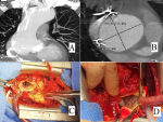
Our patient had a history of aortic valve replacement 18 years ago with a mechanical valve. After sternotomy, there is a huge pseudoaneurysm measuring 70 mm of the aortic root (A, B), totally adhering to the right atrium, which is pushed back and compressed. The aorta will be opened vertically downwards, allowing to see this enormous pseudoaneurysm encompassing the right coronary sinus (C). At this level, it exists a perforation of the aorta by an epicardial electrode and caused this pseudoaneurysm (D), which we had left in place 18 years ago to stimulate the right atrium. We finished the operation with a segment replacement by a Dacron tube.
Figure 1: A) frontal section of a thoracic CT angiography showing a pseudoaneurysm of the tubular aorta; B) thoracic CT angiography in cross-section showing a 70 mm ectasia of the aortic root; C) intraoperative view showing a huge ectasia or pseudoaneurysm encompassing the right coronary sinus; D) intraoperative view showing perforation of the aorta by an epicardial electrode
Search
This article authors
On Pubmed
On Google Scholar
Citation [Download]
Navigate this article
Similar articles in
Key words
Article metrics
Recently from the PAMJ


Authors´ services
