
Meckel´s diverticulum presenting as acute abdomen
Kiran Mastud, Yashwant Lamture
Corresponding author: Kiran Mastud, Department of General Surgery, Jawaharlal Nehru Medical College, Sawangi, Wardha, India 
Received: 13 Mar 2023 - Accepted: 04 May 2023 - Published: 23 May 2023
Domain: General surgery
Keywords: Meckel´s diverticulum, obstruction, adhesion
©Kiran Mastud et al. Pan African Medical Journal (ISSN: 1937-8688). This is an Open Access article distributed under the terms of the Creative Commons Attribution International 4.0 License (https://creativecommons.org/licenses/by/4.0/), which permits unrestricted use, distribution, and reproduction in any medium, provided the original work is properly cited.
Cite this article: Kiran Mastud et al. Meckel´s diverticulum presenting as acute abdomen. Pan African Medical Journal. 2023;45:54. [doi: 10.11604/pamj.2023.45.54.39639]
Available online at: https://www.panafrican-med-journal.com//content/article/45/54/full
Images in clinical medicine 

Meckel´s diverticulum presenting as acute abdomen
Meckel´s diverticulum presenting as acute abdomen
![]() Kiran Mastud1,&,
Kiran Mastud1,&, ![]() Yashwant Lamture1
Yashwant Lamture1
&Corresponding author

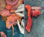
A 34-year-old female presented to us in the emergency room with acute abdomen since 2 days. Patient had tachycardia, fever, persistent vomiting, and complaint of not being able to pass stool for 2 days and flatus since 1 day. Per abdomen examination revealed a distended, tender abdomen with absent bowel sounds in all quadrants. X-ray erect abdomen was done which revealed multiple air fluid levels (more than 6) confirming the diagnosis of acute intestinal obstruction. Patient underwent an emergency exploratory laparotomy and was found to have a transition point from a mesodiverticular adhesion causing proximal dilatation of small bowel loop. A diagnosis of small bowel obstruction due to Meckel's diverticulum was made. Adhesion band was released, resection of Meckel's diverticulum and functional end-to-end anastomosis was performed. Meckel's diverticulum is a true diverticulum, containing all layers of the small bowel wall. It is failure of the vitelline duct to obliterate completely, which is usually located on the antimesenteric border of the ileum. It occurs in 2% of the general population and majority of patients remain asymptomatic. When it presents symptomatically it causes painless gastrointestinal bleeding. Nevertheless, in rare instances, it can cause acute intestinal obstruction as it did in our case. The average length of a Meckel´s diverticulum is 3 cm, with 90% ranging between 1 cm and 10 cm and the longest being 100 cm. Our case presents a large Meckel's diverticulum and the size of diverticulum was 4x2cm.
Figure 1: A,B) intraoperative and postoperative picture of Meckel´s diverticulum
Search
This article authors
On Pubmed
On Google Scholar
Citation [Download]
Navigate this article
Similar articles in
Key words
Article metrics
PlumX Metrics
Meckel´s diverticulum presenting as acute abdomenRecently from the PAMJ


Authors´ services
