
Squamous cell carcinoma in kidney with chronic pyelonephritis and pyelonephrosis: a rare case
Pragyamita Datta
Corresponding author: Pragyamita Datta, Department of Pathology, Jawaharlal Nehru Medical College (DMIHER), Sawangi, Wardha, Maharashtra, India 
Received: 28 Jan 2023 - Accepted: 20 Apr 2023 - Published: 15 May 2023
Domain: Laboratory medicine
Keywords: Calculus, kidney, pyelonephritis, squamous cell carcinoma
©Pragyamita Datta et al. Pan African Medical Journal (ISSN: 1937-8688). This is an Open Access article distributed under the terms of the Creative Commons Attribution International 4.0 License (https://creativecommons.org/licenses/by/4.0/), which permits unrestricted use, distribution, and reproduction in any medium, provided the original work is properly cited.
Cite this article: Pragyamita Datta et al. Squamous cell carcinoma in kidney with chronic pyelonephritis and pyelonephrosis: a rare case. Pan African Medical Journal. 2023;45:31. [doi: 10.11604/pamj.2023.45.31.39117]
Available online at: https://www.panafrican-med-journal.com//content/article/45/31/full
Images in clinical medicine 

Squamous cell carcinoma in kidney with chronic pyelonephritis and pyelonephrosis: a rare case
Squamous cell carcinoma in kidney with chronic pyelonephritis and pyelonephrosis: a rare case
Pragyamita Datta1,&
&Corresponding author

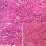
Primary squamous cell carcinoma contributes around 0.5 to 15% of all urothelial malignancy. The predisposing factor and causative agents include renal calculi, infection and endogenous and exogenous chemicals, hormonal imbalance and vitamin A deficiency. A 56-year-old female presented with right flank pain on and off with fever for the last one-week duration, imaging study revealed dilated calyx containing pus material, one focus showing a small whitish nodule. On gross examination, there was a complete loss of architecture, and one portion of calyx showed a friable whitish area and one small nodule measuring 1 x 0.5 cm. Microscopically showing an irregular nest with sheets of malignant squamous cells, differentiating into keratin pearls and background is traversed stromal invasion.
Figure 1: A, B, C) microscopically showing irregular nest and malignant cells arranged in sheets; squamous differentiation showing keratin pearls and background is traversed by stromal invasion
Search
This article authors
On Pubmed
On Google Scholar
Citation [Download]
Navigate this article
Similar articles in
Key words
Article metrics
Recently from the PAMJ


Authors´ services
