
A midline abdominal incision: can it be that dangerous? A huge umbilical vein in a cirrhotic patient
Mohamed Al Amine El Mouden, Anas Ahllat
Corresponding author: Mohamed Al Amine El Mouden, Department of General Surgery, University Hospital Center, Tangier, Morocco 
Received: 11 Nov 2022 - Accepted: 02 Dec 2022 - Published: 06 Dec 2022
Domain: General surgery
Keywords: Umbilical vein, portal hypertension, midline abdominal incision
©Mohamed Al Amine El Mouden et al. Pan African Medical Journal (ISSN: 1937-8688). This is an Open Access article distributed under the terms of the Creative Commons Attribution International 4.0 License (https://creativecommons.org/licenses/by/4.0/), which permits unrestricted use, distribution, and reproduction in any medium, provided the original work is properly cited.
Cite this article: Mohamed Al Amine El Mouden et al. A midline abdominal incision: can it be that dangerous? A huge umbilical vein in a cirrhotic patient. Pan African Medical Journal. 2022;43:178. [doi: 10.11604/pamj.2022.43.178.38187]
Available online at: https://www.panafrican-med-journal.com//content/article/43/178/full
Images in clinical medicine 

A midline abdominal incision: can it be that dangerous? A huge umbilical vein in a cirrhotic patient
A midline abdominal incision: can it be that dangerous? A huge umbilical vein in a cirrhotic patient
Mohamed Al Amine El Mouden1,&, Anas Ahllat1
&Corresponding author

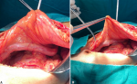
The umbilical vein transport oxygenated blood from the placenta to the fetus. It travels through the umbilical cord, which disappears at birth. It divides in two, one part going to the liver, the other one flowing into the inferior vena cava through the ductus venosus. During labor, uterine contractions cause an increase in fetal arterial pressure, which results in a decrease in the flow of para umbilical veins. A decrease in intrahepatic blood flow in the case of liver cirrhosis increases resistance in the portal vein, resulting in portal hypertension. In order to combat portal hypertension, portosystemic shunts develop in four directions: superior (left gastric vein), inferior (via hemorrhoidal veins), posterior (left renal vein), and anterior with repermeabilization of the umbilical vein and abdominal wall veins giving Cruveilhier-Baumgarten syndrome. We report the case of a 41-year-old patient followed for a hepatic cirrhosis without obvious etiology classified as child A with portal hypertension responsible for an important splenomegaly and a hypersplenism associated with a repermeabilization of the umbilical vein, operated in our general surgery department at the University Hospital of Tangier, for a non-metastatic neuroendocrine carcinoma of the gastric antrum having benefited from an antrectomy with lymph node dissection. It should be noted that the incision was in the midline above the umbilicus strict and very careful because of the risk of damaging the umbilical vein which was of large caliber and can cause a cataclysmic and uncontrollable hemorrhage. We present the impressive image of the umbilical vein in this patient.
Figure 1: A) midline abdominal incision with visualization of a huge umbilical vein; B) midline abdominal incision respecting the umbilicus
Search
This article authors
On Pubmed
On Google Scholar
Citation [Download]
Navigate this article
Similar articles in
Key words
Tables and figures
Article metrics
Recently from the PAMJ


Authors´ services
