
Unruptured myelomeningocele: a rare clinical image
Lalmalsawmi Khiangte, Pratibha Wankhede
Corresponding author: Lalmalsawmi Khiangte, Department of Community Health Nursing, Smt Radhikabai meghe memorial college of nursing, Datta Meghe Institute of Medical Sciences, Sawangi (meghe), Wardha, Maharashtra, India 
Received: 03 Nov 2022 - Accepted: 13 Nov 2022 - Published: 05 Dec 2022
Domain: Nursing education
Keywords: Neurosurgery, diagnosis, neural tube, myelomeningocele, sac
©Lalmalsawmi Khiangte et al. Pan African Medical Journal (ISSN: 1937-8688). This is an Open Access article distributed under the terms of the Creative Commons Attribution International 4.0 License (https://creativecommons.org/licenses/by/4.0/), which permits unrestricted use, distribution, and reproduction in any medium, provided the original work is properly cited.
Cite this article: Lalmalsawmi Khiangte et al. Unruptured myelomeningocele: a rare clinical image. Pan African Medical Journal. 2022;43:171. [doi: 10.11604/pamj.2022.43.171.38082]
Available online at: https://www.panafrican-med-journal.com//content/article/43/171/full
Images in clinical medicine 

Unruptured myelomeningocele: a rare clinical image
Unruptured myelomeningocele: a rare clinical image
Lalmalsawmi Khiangte1,&, Pratibha Wankhede1
&Corresponding author

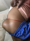
Incomplete closure of the spinal neural tube during the first month of pregnancy usually causes myelomeningocele to develop during embryonic development. It eventually results in exposed meninges or neural tissue at the level of the damaged vertebra, along with a fluid-filled sac that protrudes. Because the spinal cord does not close, many of the axons of nerves are exposed, resulting in damage to the cord as the pregnancy continues. The prognosis is frequently worse if detected late or is not treated because it can cause terrible morbidity and several impairments such as traumatic birth. An 8-year-old male child was brought to the outpatient department with complaint of swelling on the lower back which is present since birth and is increasing, which was about 15x10 cm. Physical examination was performed by the physician and the finding shows that swelling was soft with dimple and no discharge. Hence, after physical examination, the patient was diagnosed as unruptured myelomeningocele, and was referred to pediatric department for further management.
Figure 1: swelling over the lower back
Search
This article authors
On Pubmed
On Google Scholar
Citation [Download]
Navigate this article
Similar articles in
Key words
Tables and figures
Article metrics
PlumX Metrics
Unruptured myelomeningocele: a rare clinical imageRecently from the PAMJ


Authors´ services
