
Closed spinal dysraphism-lipomyelocele in an 18-year-old male
Nikita Hitesh Seth, Ragini Dadgal
Corresponding author: Ragini Dadgal, Ravi Nair Physiotherapy College, Datta Meghe Institute of Medical Sciences, Sawangi, Wardha, Maharashtra, India 
Received: 19 Feb 2022 - Accepted: 09 May 2022 - Published: 23 May 2022
Domain: Radiology
Keywords: Lipomyelocele, physical therapy, unhealing foot ulcers
©Nikita Hitesh Seth et al. Pan African Medical Journal (ISSN: 1937-8688). This is an Open Access article distributed under the terms of the Creative Commons Attribution International 4.0 License (https://creativecommons.org/licenses/by/4.0/), which permits unrestricted use, distribution, and reproduction in any medium, provided the original work is properly cited.
Cite this article: Nikita Hitesh Seth et al. Closed spinal dysraphism-lipomyelocele in an 18-year-old male. Pan African Medical Journal. 2022;42:60. [doi: 10.11604/pamj.2022.42.60.33893]
Available online at: https://www.panafrican-med-journal.com//content/article/42/60/full
Images in clinical medicine 

Closed spinal dysraphism-lipomyelocele in an 18-year-old male
Closed spinal dysraphism-lipomyelocele in an 18-year-old male
![]() Nikita Hitesh Seth1,
Nikita Hitesh Seth1, ![]() Ragini Dadgal1,&
Ragini Dadgal1,&
&Corresponding author

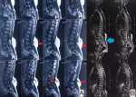
We are reporting a magnetic resonance imaging (MRI) finding of an 18-year-old male not a known case of any chronic medical illness presented with chief complaints of swelling over the lumbosacral region since birth with sensory loss over bilateral lower limb for 2 months. He also complained of unhealing ulcers over the dorsum of the great toe. Magnetic Resonance Imaging (MRI) findings revealed T2 hyperintensive mass (red arrow) of subcutaneous soft tissue in the posterior lumbosacral region extending into the spinal canal. Posterior spinal defect at the L3-L4 level with tethering of cord was noted, suggestive of Lipomyelocele. Following the removal of the Lipomyelocele and detethering of the chord, the patient suffered bladder dysfunction and right foot drop. For additional treatment, he was referred to a physiotherapist. He had weakness in his lower limbs ( Medical Research Council (MRC) grade 3), a foot drop on the right side, and considerable muscular loss on physical examination. Sensations were affected on both sides. Bladder training, sensory re-education, lower limb muscular strengthening, and foot drop management were all part of the physiotherapy rehabilitation. Foot drop was treated with an ankle-foot orthosis and dorsiflexion reflex stimulation, with an emphasis on activities in a weight-bearing position. It has been discovered that a multidisciplinary strategy involving medical, surgical, and physical therapy improves treatment outcomes.
Figure 1: A) red arrow indicates lipomyelocele at lumbosacral region; B) post-operative MRI, blue arrow indicates thickened filum terminale and tethering at L2-L3 level
Search
This article authors
On Pubmed
On Google Scholar
Citation [Download]
Navigate this article
Similar articles in
Key words
Tables and figures
Article metrics
Recently from the PAMJ


Authors´ services
