
Osteochondroma of rib
Prateek Upadhyay
Corresponding author: Prateek Upadhyay, Department of Orthopaedics, Jawaharlal Nehru Medical College, Datta Meghe Institute of Medical Sciences (Deemed to be University), Wardha, Maharashtra, India 
Received: 30 Apr 2022 - Accepted: 13 May 2022 - Published: 23 May 2022
Domain: Orthopedic surgery
Keywords: Ostechondroma, rib, vetebrae
©Prateek Upadhyay et al. Pan African Medical Journal (ISSN: 1937-8688). This is an Open Access article distributed under the terms of the Creative Commons Attribution International 4.0 License (https://creativecommons.org/licenses/by/4.0/), which permits unrestricted use, distribution, and reproduction in any medium, provided the original work is properly cited.
Cite this article: Prateek Upadhyay et al. Osteochondroma of rib. Pan African Medical Journal. 2022;42:59. [doi: 10.11604/pamj.2022.42.59.35217]
Available online at: https://www.panafrican-med-journal.com//content/article/42/59/full
Images in clinical medicine 

Osteochondroma of rib
Osteochondroma of rib
![]() Prateek Upadhyay1,&
Prateek Upadhyay1,&
&Corresponding author

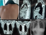
Osteochondroma is also known as exostosis and is a benign bone tumour. It commonly presents in the first 3 decades of life with a male preponderance. The most common site for the development of these tumours is the metaphysis of long bones, with almost 30% of cases originating from the distal metaphysis of the femur. Origin from flat bones like ilium and scapula is rare, while origin from vertebrae and ribs is unheard of. A 21-year-old female presented with a history of swelling over the left upper back since 2 years, pain in the left upper back and shoulder with tingling since 4 months. On examination, a 3 cm by 3 cm, hard, immobile mass was palpable in the left paravertebral region of the 4th intercostal space. X-ray and computed tomography (CT) scan of the thorax revealed two irregular lesions arising from the neck of the 5th rib posteriorly and a Fine Needle Aspiration Cytology (FNAC) revealed the lesion to be of chondroid matrix origin, consisting of normal chondrocytes. Through a posterior approach, two pedunculated tumours of size 3.5 x 2 x 1�cm and 2 x 2 x 1�cm, along with the costovertebral junction of the 5th rib were resected. Histopathological examination confirmed the mass to be an osteochondroma of the rib. On 6 months follow up, the patient is stable, has no pain or tingling and there is no evidence of recurrence of the tumour.
Figure 1: A) clinical picture of the swelling in the left 4th intercostal region; B) sagittal section of CT thorax showing the irregular mass between the 4th and 5th rib; C) coronal section of CT thorax showing the irregular mass between the 4th and 5th rib; D) post-operative X-ray with resected mass and the costovertebral junction of the 5th rib
Search
This article authors
On Pubmed
On Google Scholar
Citation [Download]
Navigate this article
Similar articles in
Key words
Article metrics
PlumX Metrics
Osteochondroma of ribRecently from the PAMJ


Authors´ services
