
Gastroschisis, a congenital anterior abdominal wall defect: a rare clinical image
Ashna Gledina, Seema Singh
Corresponding author: Ashna Gledina, Department of Medical Surgical Nursing, Smt. Radhikabai Meghe Memorial College of Nursing, Datta Meghe Institute of Medical Sciences, Sawangi, Wardha, Maharashtra, India 
Received: 17 Jul 2022 - Accepted: 19 Jul 2022 - Published: 22 Aug 2022
Domain: Pediatric gastroenterology
Keywords: Gastroschisis, congenital malformation, birth defect, abdominal wall defect
©Ashna Gledina et al. Pan African Medical Journal (ISSN: 1937-8688). This is an Open Access article distributed under the terms of the Creative Commons Attribution International 4.0 License (https://creativecommons.org/licenses/by/4.0/), which permits unrestricted use, distribution, and reproduction in any medium, provided the original work is properly cited.
Cite this article: Ashna Gledina et al. Gastroschisis, a congenital anterior abdominal wall defect: a rare clinical image. Pan African Medical Journal. 2022;42:298. [doi: 10.11604/pamj.2022.42.298.36378]
Available online at: https://www.panafrican-med-journal.com//content/article/42/298/full
Images in clinical medicine 

Gastroschisis, a congenital anterior abdominal wall defect: a rare clinical image
Gastroschisis, a congenital anterior abdominal wall defect: a rare clinical image
Ashna Gledina1,&, Seema Singh1
&Corresponding author

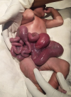
Gastroschisis is a relatively rare birth defect in which the baby´s intestines (stomach, large or small intestines) extends outside of the abdominal wall or exit their body from a 2 to 5 cm hole, most often on the right side beside their belly button during fetal development. It occurs in about 1 in every 2,000 babies. We report a case of a large gastroschisis containing intestinal loop. A 29-year-old, primigravida, was referred to the centre for childbirth. Antenatally, at 24th weeks the fetus was diagnosed with a congenital malformation of the anterior abdominal wall. The ultrasound at 30 weeks, confirmed the diagnosis. At 37 plus weeks, she was taken for emergency caesarean section. A male child was born with the confirmation of presence of gastroschisis. Birth weight 3.2 Kg, height 54 cm with appearance, pulse, grimace, activity, and respiration (APGAR) score 8/9. At birth gastroschisis contained intestinal loop. Newborn was referred to neonatal intensive for further management.
Figure 1: loops of the baby´s intestines (stomach, large and small intestines) extended outside the abdomen wall
Search
This article authors
On Pubmed
On Google Scholar
Citation [Download]
Navigate this article
Similar articles in
Key words
Tables and figures
Article metrics
Recently from the PAMJ


Authors´ services
