
Pityriasis rubra pilaris with rickets: a rare clinical image
Sangita Shende, Ranjana Sharma
Corresponding author: Sangita Shende, Florence Nightingale Training College of Nursing, Datta Meghe Institute of Medical Science University of Delhi (DU), Sawangi Meghe, Wardha, Maharashtra, India 
Received: 02 Jul 2022 - Accepted: 01 Aug 2022 - Published: 16 Aug 2022
Domain: Dermatology
Keywords: Pityriasis rubra pilaris, systemic therapy, papulosquamous, Vitamin D, deficiency
©Sangita Shende et al. Pan African Medical Journal (ISSN: 1937-8688). This is an Open Access article distributed under the terms of the Creative Commons Attribution International 4.0 License (https://creativecommons.org/licenses/by/4.0/), which permits unrestricted use, distribution, and reproduction in any medium, provided the original work is properly cited.
Cite this article: Sangita Shende et al. Pityriasis rubra pilaris with rickets: a rare clinical image. Pan African Medical Journal. 2022;42:285. [doi: 10.11604/pamj.2022.42.285.36169]
Available online at: https://www.panafrican-med-journal.com//content/article/42/285/full
Images in clinical medicine 

Pityriasis rubra pilaris with rickets: a rare clinical image
Pityriasis rubra pilaris with rickets: a rare clinical image
Sangita Shende1,&, ![]() Ranjana Sharma2
Ranjana Sharma2
&Corresponding author

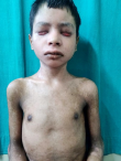
Pityriasis rubra pilaris (PRP) manifests as well-defined erythematous scaly plaques with follicular keratosis mainly over the elbows and knees. We present a case of a 15-years-old boy who comes to the dermatology department with complaints of itching, dryness, burning sensation, photosensitivity, erythematous skin with exfoliation over face and neck, hyperpigmentation over bilateral hand and trunk fissuring over bilateral feet, ectropion of bilateral lower lids from 3 months. On physical examination, the patient face had lesions and redness on the eyebrow. Skin biopsy and blood investigation revealed the received single, irregular, whitish tissue piece with skin and hair attached measuring 0.2x0.2 cm section shows histopathological features suggestive of pityriasis rubra pilaris. Ortho review call and review of investigations and X-rays advised the patient diagnosed with rickets. The patient was referred to the dermatology department for further management.
Figure 1: clinical image shows erythematous skin with exfoliation over face and neck, and hyperpigmentation over bilateral hand
Search
This article authors
On Pubmed
On Google Scholar
Citation [Download]
Navigate this article
Similar articles in
Key words
Tables and figures
Article metrics
Recently from the PAMJ


Authors´ services
