
A rare clinical image of fourth-degree bedsore
Minal Dambhare, Deeplata Mendhe
Corresponding author: Minal Dambhare, Nursing Tutor Florence Nightingale Training College of Nursing, Datta Meghe Institute of Medical Science (DU) Sawangi Meghe Wardha, Wardha, Maharashtra, India 
Received: 30 Jun 2022 - Accepted: 05 Jul 2022 - Published: 11 Aug 2022
Domain: Emergency medicine
Keywords: Decubitus ulcer, spinal injury, bedsore
©Minal Dambhare et al. Pan African Medical Journal (ISSN: 1937-8688). This is an Open Access article distributed under the terms of the Creative Commons Attribution International 4.0 License (https://creativecommons.org/licenses/by/4.0/), which permits unrestricted use, distribution, and reproduction in any medium, provided the original work is properly cited.
Cite this article: Minal Dambhare et al. A rare clinical image of fourth-degree bedsore. Pan African Medical Journal. 2022;42:271. [doi: 10.11604/pamj.2022.42.271.36133]
Available online at: https://www.panafrican-med-journal.com//content/article/42/271/full
Images in clinical medicine 

A rare clinical image of fourth-degree bedsore
A rare clinical image of fourth-degree bedsore
Minal Dambhare1,&, ![]() Deeplata Mendhe2
Deeplata Mendhe2
&Corresponding author

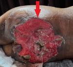
Stage four bedsores are the most severe form of bedsores, also called pressure sores, pressure ulcers, or decubitus ulcers. A stage four bedsore is a deep wound reaching muscles, ligaments, or bones. They often cause residents to suffer extreme pain, infection, and invasive surgeries. Its major etiological factors are poor circulation, Immobility due to spinal cord injury, Excessive moisture, skin irritants like urine and feces, and friction. It mainly occurs in men and women, both aged 20 to 80 years. With early diagnosis and treatment, further complications can be prevented. We report a case of the 29-year-old patient; who came to the surgical intensive care unit with the complaints of swelling over the buttocks, in size of 20 x 20 x 8 cm bedsore covering both buttocks, foul-smelling he was apparently alright five years back when he noticed swelling and skin off on buttocks with itching and burning pain which was the insidious onset and progressive. Initially, the size was 20 x 20 x 8 cm, gradually increasing to the current size of 24 x 24 cm. He also complains of pain over the buttock, a throbbing type of pain. A blood investigation and skin biopsy were done, and the patient was referred to the surgical intensive care unit for further management.
Figure 1: deep wound with redness of the skin
Search
This article authors
On Pubmed
On Google Scholar
Citation [Download]
Navigate this article
Similar articles in
Key words
Tables and figures
Article metrics
PlumX Metrics
A rare clinical image of fourth-degree bedsoreRecently from the PAMJ


Authors´ services
