
A traumatic injury leading the lipoma of neck: a rare image
Heena Hussain Pathan, Sanskarsingh Vijaysingh Banafar
Corresponding author: Heena Hussain Pathan, Department of Community Physiotherapy, Ravi Nair Physiotherapy College, Datta Meghe Institute of Medical Sciences, Wardha, Maharashtra, India 
Received: 21 Jun 2022 - Accepted: 04 Jul 2022 - Published: 26 Jul 2022
Domain: Laboratory medicine,Physical medicine and rehabilitation�or Physiatry,Urgent Care Medicine
Keywords: Lipoma, hypertension, benign tumor
©Heena Hussain Pathan et al. Pan African Medical Journal (ISSN: 1937-8688). This is an Open Access article distributed under the terms of the Creative Commons Attribution International 4.0 License (https://creativecommons.org/licenses/by/4.0/), which permits unrestricted use, distribution, and reproduction in any medium, provided the original work is properly cited.
Cite this article: Heena Hussain Pathan et al. A traumatic injury leading the lipoma of neck: a rare image. Pan African Medical Journal. 2022;42:234. [doi: 10.11604/pamj.2022.42.234.36014]
Available online at: https://www.panafrican-med-journal.com//content/article/42/234/full
Images in clinical medicine 

A traumatic injury leading the lipoma of neck: a rare image
A traumatic injury leading the lipoma of neck: a rare image
![]() Heena Hussain Pathan1,&,
Heena Hussain Pathan1,&, ![]() Sanskarsingh Vijaysingh Banafar2
Sanskarsingh Vijaysingh Banafar2
&Corresponding author

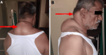
A 58 years old male visited our hospital with the complaint of swelling over the posterior aspect of the neck after sustaining a trauma as a result of a fall of a cement slab over him 5 years back. The patient is under a combination of metoprolol and telmisartan medication for the rectification of hypertension for 10 years. The patient got admitted to the hospital for the above complaint and has undergone a histopathology report, MRI, and USG investigation. In the histopathology report, we received a single container labelled as a resected specimen of large lipoma. On the cut section, a solid, yellowish, homogenous area was identified. MRI investigation reported a lesion over the nape of the neck size measuring from 25x20x8cm in size. USG report stated a lesion over the nape of the neck and occipital region with the dimensions of the left side as 53x25mm approximately, right side as 58x31.5�mm approximately. After both MRI and USG investigation, the patient was diagnosed with lipoma over the nape of the neck and occipital region. The patient had gone through two separate operations in a spare of 11 days. Complete excision of Excessive fibrofatty tissue size 10x15 cm lipoma was done. Flap was approximated and romovac drian no 14 placed. Closure was done with ethilon2-0 ac. PPd debridement was done with secondary suturing. Hemostasis was confirmed and Patient stable. Shifted to ward and got discharged after 1 week with prescribed medications and physiotherapy exercise.
Figure 1: A) dorsal view showing swelling over the nape of the neck; B) lateral view showing swelling over the occipital region
Search
This article authors
On Pubmed
On Google Scholar
Citation [Download]
Navigate this article
Similar articles in
Key words
Tables and figures
Article metrics
Recently from the PAMJ


Authors´ services
