
Lumbar rachischisis associated with diastematomyelia: prenatal diagnosis and fetopathological examination
Mahdi Farhati, Abir Karoui
Corresponding author: Mahdi Farhati, Obstetrics and Gynecology Department “C”, Tunis Maternity and Neonatology Center, Tunis, Tunisia 
Received: 27 May 2022 - Accepted: 04 Jun 2022 - Published: 23 Jun 2022
Domain: Radiology,Obstetrics and gynecology
Keywords: Prenatal diagnosis, rachischisis, spina bifida, diastematomyelia, sonography
©Mahdi Farhati et al. Pan African Medical Journal (ISSN: 1937-8688). This is an Open Access article distributed under the terms of the Creative Commons Attribution International 4.0 License (https://creativecommons.org/licenses/by/4.0/), which permits unrestricted use, distribution, and reproduction in any medium, provided the original work is properly cited.
Cite this article: Mahdi Farhati et al. Lumbar rachischisis associated with diastematomyelia: prenatal diagnosis and fetopathological examination. Pan African Medical Journal. 2022;42:146. [doi: 10.11604/pamj.2022.42.146.35644]
Available online at: https://www.panafrican-med-journal.com//content/article/42/146/full
Images in clinical medicine 

Lumbar rachischisis associated with diastematomyelia: prenatal diagnosis and fetopathological examination
Lumbar rachischisis associated with diastematomyelia: prenatal diagnosis and fetopathological examination
![]() Mahdi Farhati1,&,
Mahdi Farhati1,&, ![]() Abir Karoui1
Abir Karoui1
&Corresponding author

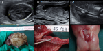
We report the case of a 29-year-old female patient who presented for a routine ultrasound scan at 17 weeks of gestation. Sonography showed, on an axial view of the fetal brain, flattening of the frontal bones resulting in a lemon like deformity of the cephalic pole (A). Biparietal diameter and head circumference were below 3rd percentile (A). The examination of lumbar spine showed vertebral and skin defect on a sagittal view (B). The diagnosis of Chiari II malformation was suspected. An additional midline echogenic focus (B, C) attracted our attention. Sonography showed widening of the spinal canal in the coronal view (C). Associated diastematomyelia was suspected. Magnetic resonance imaging (MRI) confirmed the presence of lumbar rachischisis associated with diastematomyelia. The couple decided to interrupt the pregnancy. Fetopathological examination confirmed the abnormalities described on ultrasound (A, B, C) and showed an associated ventricular septal defect.
Figure 1: (A, B, C) prenatal sonography findings and fetopathology examination
Search
This article authors
On Pubmed
On Google Scholar
Citation [Download]
Navigate this article
Similar articles in
Key words
Article metrics
Recently from the PAMJ


Authors´ services
