
Giant urinary bladder calculi in a 60-year-old man: a case report
Faisal Ahmed, Qasem Alyhari, Saleh Al-wageeh, Fawaz Mohammed
Corresponding author: Faisal Ahmed, Urology Research Center, Al-Thora General Hospital, Department of Urology, School of Medicine, Ibb University of Medical Science, Ibb, Yemen 
Received: 06 Jan 2022 - Accepted: 13 Jan 2022 - Published: 27 Jan 2022
Domain: Urology
Keywords: Bladder stone, cystolithotomy, giant, case report
©Faisal Ahmed et al. Pan African Medical Journal (ISSN: 1937-8688). This is an Open Access article distributed under the terms of the Creative Commons Attribution International 4.0 License (https://creativecommons.org/licenses/by/4.0/), which permits unrestricted use, distribution, and reproduction in any medium, provided the original work is properly cited.
Cite this article: Faisal Ahmed et al. Giant urinary bladder calculi in a 60-year-old man: a case report. Pan African Medical Journal. 2022;41:78. [doi: 10.11604/pamj.2022.41.78.33131]
Available online at: https://www.panafrican-med-journal.com//content/article/41/78/full
Case report 

Giant urinary bladder calculi in a 60-year-old man: a case report
Giant urinary bladder calculi in a 60-year-old man: a case report
![]() Faisal Ahmed1,&, Qasem Alyhari2,
Faisal Ahmed1,&, Qasem Alyhari2, ![]() Saleh Al-wageeh2,
Saleh Al-wageeh2, ![]() Fawaz Mohammed3
Fawaz Mohammed3
&Corresponding author
Urinary bladder calculi comprise 5% of all urinary tract calculi. Giant bladder calculi are defined as a stone more than 100g in weight. However, giant bladder calculus weighted more than 500g is rare in current practice. We present a 60-year-old man who presented with dysuria, difficulty in urination, and suprapubic pain started four years ago. The plain radiology image showed big intravesical caliculi measured about 10x9cm. The calculi was removed via open cystolithotomy without postoperative complication. The caliculi weighed 750g. In conclusion, the main goal of treatment is to remove the calculi and relieve the accompanying symptoms.

Giant urinary bladder calculi are defined as a stone more than 100g in weight, a rare condition in current practice [1]. Urinary tract infection, urethral stricture, benign prostatic hyperplasia, and intravesical foreign body encrustation are the most common causes of urinary bladder calculi formation [2]. Bladder calculi present in various ways, ranging from entirely asymptomatic to dysuria, suprapubic pain, hematuria, and urine retention [3]. A few published reports regarding giant bladder stones weighed more than 500g [4]. Here we present a case of giant bladder calculi in a 60-year-old man. The manifestations, diagnosis, and treatment are discussed.

Patient information: a 60-year-old Yemeni man, illiterate, presented to our urology department at Althora general hospital in September 2021 with a chief complaint of dysuria, difficulty in urination, and suprapubic pain started four years ago. There is no history of abdominal trauma, neurological disease, or urologic disease such as urinary tract stones.
Clinical findings: regarding the physical examination, the patient had severe, nonradiated suprapubic pain associated with mild tenderness and detectable enlarged and nonmobile suprapubic mass in palpation.
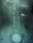
Diagnostic assessment: blood tests revealed a total white blood cell count of 12x10³/ml with moderate leukocytosis and hemoglobin: 14.4g/dl, blood urea nitrogen: 14mg/dl, and creatinine: 1.1mg/dl. Urine analysis showed microscopic hematuria (15-20 RBCs/HPF) and many pass cells (20 WBCs/HPF). The abdominal X-ray showed giant intravesical calculus measured 10x9cm (Figure 1). The ultrasonography showed moderate hydronephrosis in both kidneys, mild bladder wall thickness, giant intravesical calculi, and mild prostate enlargement.
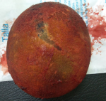
Therapeutic interventions: the antibiotic (Ceftriaxone g every 12hours for three days) was started to control urinary tract infection. Then, the patient was admitted to elective surgery. Via spinal anesthesia in the supine position, open cystolithotomy was made with a lower umbilical midline incision, the bladder was opened, and large calculus adhered to the bladder wall and measured about 10x9x5cm was observed and removed. Three ways urethral catheter was inserted, and the bladder and skin were finally closed. The calculi weighed about 750g (Figure 2).
Follow-up and outcome: postoperative hospital course was uneventful. The patient was discharged with an oral antibiotic on the third postoperative day, and the catheter was removed on the seventh postoperative day. Within three months of follow-up, the patient remained symptom-free.
Patient perspective: the patient was happy with the successful outcome of the surgery.
Informed consent: written informed consent was obtained from the patient for participation in our study.
Giant urinary bladder calculi is a rare condition and defined stone more than 100g in weight [1]. It is more frequent in males than females [2]. Giant urinary bladder calculi most often occur with other pathologic bladder conditions such as urinary retention, urethral stricture, urinary tract infection, prolonged catheterization, neurogenic bladder, and the existence of the foreign body [5].
The pathophysiology of giant bladder calculi can develop from a nidus of infected material or single ureteric calculus with a progressive layer-wise accumulation of calcified matrix associated with factors causing urinary stasis such as bladder outlet obstruction [2]. Factors such as low educational level, low socioeconomic, dry climate, dietary habits, and high exposure to the sun are associated with a high incidence of urinary tract calculi [6]. Our patient was illiterate, worked outdoor with high exposure to the sun.
The clinical manifestations of giant bladder calculi range from entirely asymptomatic to acute urinary retention [4]. Our patient was suffered from dysuria, difficulty in urination, and suprapubic pain. The vast majority of bladder calculi are radiopaque and can be identified with a plain radiograph. Other useful radiologic images are ultrasonography, computed tomography scan, magnetic resonance imaging, and intravenous pyelography [7,8].
There are several surgical options for bladder stone, including open cystolithotomy, extracorporeal fragmentation, percutaneous endoscopic cystolitholapexy, and cystolitholapexy. Open cystolithotomy has been recommended as the best treatment option for large bladder calculi [3,4]. Similarly, our patient was undergoing open cystolithotomy without complication.
Few recently published reports with giant bladder calculi weighted more than 500g such as Ma and associations who reported a bladder calculi with 1048g in weight [4], Shrestha et al. reported a bladder calculi with 950g in weight [9], Nugroho and associations reported a bladder calculi with 832g in weight [3], and the recent report by Pattiiha et al. reported a bladder calculi with 500g in weight [6]. Similarly, our patient had giant bladder calculus that weighed 750g.
Giant urinary bladder calculi with more than 500g in weight are rare in current urologic practice. The radiologic diagnostic methods of bladder calculi include ultrasonography, plain radiography, and computed tomography scan. Open cystolithotomy is our patient's best surgical option in giant bladder calculi.
The authors declare no competing interests.
Patient management: FA. Data collection, manuscript drafting, and revision: SA, FM, and QA. All authors read and approved the final version of the manuscript.
The authors would like to thank the General Manager of Althora General Hospital, Ibb, Yemen, Dr. Abdulghani Ghabisha, for editorial assistance.
Figure 1: giant bladder stone in plain radiography
Figure 2: a 10x9cm bladder stone removed by open cystolithotomy
- Becher RM, Tolia BM, Newman HR. Giant vesical calculus. Jama. 1978 May 26;239(21):2272-3. Epub 1978/05/26. PubMed | Google Scholar
- Vidhyarthy AK, Hameed T, Lal R, Kumar A, Sahni S, Mendoza N. Giant Bladder Calculus in an Adult- A Persistent Problem in the Developing World: A Case Report. Clin Pract Cases Emerg Med. 2020 Nov;4(4):544-7. Epub 2020/11/21. PubMed | Google Scholar
- Nugroho EA, Junita D, Wijaya YH. Giant bladder stone with history of recurrence urinary tract infections: A rare case. Urol Case Rep. 2019 Sep;26:100945. Epub 2019/07/06. PubMed | Google Scholar
- Ma C, Lu B, Sun E. Giant bladder stone in a male patient: A case report. Medicine (Baltimore). 2016 Jul;95(30):e4323. PubMed PMID: 27472711. Epub 2016/07/30. PubMed | Google Scholar
- Hizli F, Yilmaz E. A giant bladder struvite stone in an adolescent boy. Urol Res. 2012 Jun;40(3):273-4. Epub 2011/12/08. PubMed | Google Scholar
- Pattiiha AM, Hadi AF, Rokhimah S, Hafiq HM. Giant bladder uric acid stone with a history of prolonged sun exposure and high protein diet in North Moluccas: Case series. Int J Surg Case Rep. 2020;73:328-31. PubMed | Google Scholar
- Ahmed F, Askarpour MR, Eslahi A, Nikbakht HA, Jafari SH, Hassanpour A et al. The role of ultrasonography in detecting urinary tract calculi compared to CT scan. Res Rep Urol. 2018;10:199-203. Epub 2018/12/05. PubMed | Google Scholar
- Hammad FT, Kaya M, Kazim E. Bladder calculi: did the clinical picture change? Urology. 2006 Jun;67(6):1154-8. Epub 2006/06/13. PubMed | Google Scholar
- Shrestha N, Zhou L, Hu CH. Extraction of giant bladder calcium oxalate stone: A case report. Int J Surg Case Rep. 2020;68:151-3. Epub 2020/03/0. PubMed | Google Scholar
Search
This article authors
On Pubmed
On Google Scholar
Citation [Download]
Navigate this article
Similar articles in
Key words
Tables and figures
Article metrics
Recently from the PAMJ


Authors´ services
