
Caroli´s disease incidentally discovered in a 16-years-old female: a case report
Abdullatif Almohtadi, Faisal Ahmed, Fawaz Mohammed, Morad Sanhan, Abdulghani Ghabisha, Lina Al-moliki
Corresponding author: Faisal Ahmed, Urology Research Center, Al-Thora General Hospital, Department of Urology, School of Medicine, Ibb University of Medical Science, Ibb, Yemen 
Received: 02 Mar 2022 - Accepted: 08 Mar 2022 - Published: 14 Mar 2022
Domain: Radiology,Hepatology
Keywords: Caroli´s disease, abdominal pain, cholangitis, case report
©Abdullatif Almohtadi et al. Pan African Medical Journal (ISSN: 1937-8688). This is an Open Access article distributed under the terms of the Creative Commons Attribution International 4.0 License (https://creativecommons.org/licenses/by/4.0/), which permits unrestricted use, distribution, and reproduction in any medium, provided the original work is properly cited.
Cite this article: Abdullatif Almohtadi et al. Caroli´s disease incidentally discovered in a 16-years-old female: a case report. Pan African Medical Journal. 2022;41:204. [doi: 10.11604/pamj.2022.41.204.34088]
Available online at: https://www.panafrican-med-journal.com//content/article/41/204/full
Case report 

Caroli´s disease incidentally discovered in a 16-years-old female: a case report
Caroli´s disease incidentally discovered in 16-years-old female: a case report
![]() Abdullatif Almohtadi1,
Abdullatif Almohtadi1, ![]() Faisal Ahmed2,&,
Faisal Ahmed2,&, ![]() Fawaz Mohammed3, Morad Sanhan4, Abdulghani Ghabisha4, Lina Al-moliki4
Fawaz Mohammed3, Morad Sanhan4, Abdulghani Ghabisha4, Lina Al-moliki4
&Corresponding author
Caroli´s disease is a congenital hepatic disorder characterized by nonobstructive saccular or fusiform dilatation of the intrahepatic bile ducts with the absence of congenital hepatic fibrosis. Caroli´s disease is rare, with few reported cases in the literature, making it hard to distinguish from other liver abnormalities. We present a case of Caroli´s disease discovered indecently in a 16-year-old female who presented with recurrent abdominal pain and intermittent jaundice in the last three years. Abdominal Computed tomography (CT) showed mild liver enlargement with multiple cystic dilatations of the intrahepatic saccular bile ducts cystic dilatations without hepatic fibrosis. The patient was treated conservatively with ursodeoxycholic acid and antibiotic therapy and discharged with regular follow-up. In conclusion, Caroli´s disease should be considered in the differential diagnosis in patients with recurrent abdominal pain and cholangitis without risk factors or relevant history.

Caroli´s disease is a rare liver congenital malformation with a prevalence rate of less than one in one million societies with a male to female ratio of 1:1.8 [1]. The main features of Caroli´s disease are intrahepatic bile duct dilation with biliary tract involvement in the focal or multifocal direction, while the absence of congenital hepatic fibrosis distinguishes it from Caroli syndrome [2,3]. The typical symptoms of Caroli´s disease are abdominal pain, itching, jaundice, and recurrent cholangitis, which occurred due to liver insufficiency and portal hypertension [3]. It may associate with splenomegaly, abdominal ascites, edema, coagulopathy, and esophageal varices [4]. Caroli´s disease is rare, with few reported cases in the literature, making it hard to distinguish from other liver abnormalities [4]. Hence, we present a 16-year-old female presented with cholangitis and was diagnosed with Caroli´s disease by computed tomography scan. This study aims to add to current knowledge about this sporadic congenital disease.

Patient information: a 16-years-old female presented with abdominal pain, low-grade fever, and jaundice three days ago. The pain was mild and was located in the right upper quadrant. She had no history of nausea, vomiting, weight loss, changes in bowel habits, or alcohol consumption. The patient mentioned a history of chronic abdominal pain, recurrent jaundice, and repeated hospital admission due to her pain without improvement. The patient was not a smoker and there was no family history of congenital or hereditary diseases.
Clinical findings: the vital signs were stable, and the oral temperature was 37.9°C. The abdomen was mildly distended with mild tenderness in the right upper quadrant and mild hepatosplenomegaly.
Diagnostic assessment: the white blood cell count:12 �10³/ml, hemoglobin: 11 g/dl, platelets: 260�10³/ml, blood urea nitrogen: 14 mg/dl, and creatinine:1.1 mg/dl, total bilirubin: 5 mg/dl, direct bilirubin: 3.5 mg/dl, albumin: 4.3 g/dL, alkaline phosphatase: 305 U/L, aspartate aminotransferase (AST): 24 UI/mL, and alanine aminotransferase (ALT): 13 UI/ml. The viral hepatitis marker was negative. The coagulation tests: prothrombin time (PT), international normalized ratio (INR) and partial thromboplastin time (PTT) were all within the normal range.
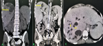
Abdominal ultrasound (US) showed multiple intrahepatic cysts associated with hepatomegaly and splenomegaly. Computed tomography (CT) scan of the abdomen showed mild enlargement of the liver with multiple multifocal hypodense intrahepatic cystic lesions in all segments and the largest one located at a posterior right upper lope, measuring about 3.6�2.8cm, and closely connected to intrahepatic biliary ducts without common bile duct dilatation or hepatic fibrosis. Mild portal and liver vein dilatations, hepatosplenomegaly, and mild abdominal ascites were observed (Figure 1).
Therapeutic interventions: the patient was newly diagnosed with Caroli´s disease, so conservative medical therapy was started. She has been treated with an intravenous antibiotic; Cefuroxime 750 mg every 8 hours for 5 days, Metronidazole 500mg every 8 hours, Ursodeoxycholic acid every 12 hours and Pantoprazole 40 mg daily.
Follow-up and outcome: after five days, the patient's general condition improved, and she was discharged with regular follow-up.
Patient perspective: at the 6-month follow-up, the patient reported that she continued taking the prescribed medications. She stated that she felt more energized and could attend school and work more frequently. She also mentioned feeling more rested, which she attributed to the absence of abdominal pain and itching.
Written informed consent: written informed consent was obtained from the patient for participation in our study.
The pathophysiology of Caroli´s disease is still unknown. However, a genetic profile seems to be autosomal recessive [4]. An unbalanced translocation between chromosomes 3 and 8 was discovered, implying that loss of distal 3p and/or gain of 8q could be pathogenetic in Caroli´s disease [5]. Possible mechanisms for cystic dilatations in Caroli´s disease are: obstruction of the hepatic artery leads to bile duct ischemia and cystic dilatation, overgrowth of the bile epithelium and connective tissue development, lack of normal molting of the border duct plates with the portal ducts and consequently in the cysts around the portal triads [6,7].
Recurrent abdominal pain in the right upper quadrant area, itching, and jaundice with recurrent bacterial cholangitis are the most manifestations of Caroli´s disease [4]. Our patient was presented with the same manifestations.
The US, abdominal CT, magnetic resonance cholangiopancreatography (MRCP), and magnetic resonance imaging (MRI) are the usual radiologic method for investigations. Despite being operator-sensitive and having low specificity, the US provides the best initial examination because it is noninvasive, simple, and inexpensive. The US findings are unusual dilation of the intrahepatic bile ducts and dilation of the extrahepatic ducts in some cases due to cholelithiasis [4,8]. MRCP is associated with reasonable precision, high predictability, and low potential complications. It is currently the optimal radiographic option. Nevertheless, it is more expensive and used only for determining the disease's nature and severity [6,9]. Abdominal CT scans with intravenous contrast enhancement may reveal central dots in this disease [4,10]. Our patient incidentally detected dilated hepatic cystic lesions on an abdominal CT scan compatible with Caroli´s disease.
There are no exact guidelines for treating Caroli´s disease due to its rarity. However, the proposed therapy should adapt to the clinical manifestations and the potential consequences of biliary abnormalities. Ursodeoxycholic acid is the chosen treatment option in a patient with mild symptoms. Ursodeoxycholic acid has three significant mechanisms of action: the first protects cholangiocytes from the cytotoxic activity of hydrophobic bile acids. The second mechanism is the stimulation of hepatobiliary secretion. The last mechanism is hepatocyte protection against bile acid-induced apoptosis [11]. In patients with symptoms of cholecystitis, instead of ursodeoxycholic acid, a proper antibiotic therapy should be started [3]. The main indication for surgical interventions in Caroli´s disease are biliary obstruction, abscess formation, and bile duct stones. Additionally, patients with an advanced disease stage may qualify for a liver transplant [3,4]. Our patient had mild symptoms treated with ursodeoxycholic acid and proper antibiotics for symptoms of cholangitis.
The differential diagnosis of Caroli´s disease is primary sclerosing cholangitis, recurrent pyogenic cholangitis, polycystic liver disease, choledochal cysts, biliary papillomatosis, and the Von Meyenburg complex [12]. The Von Meyenburg complex usually does not cause symptoms or disturbances in liver functions. MRCP is the optimal choice for its diagnosis, characterized by multiple small-size cysts less than 15 mm and does not communicate with the biliary tree [13].
Caroli´s disease is a rare congenital malformation of the intahepatic bile ducts. Despite its low prevalence, Caroli´s disease should be considered in the differential diagnosis in patients with recurrent abdominal pain and cholangitis without risk factors or relevant history. Radiologic investigations such as the US, abdominal CT scans, and MRCP are helpful to confirm the diagnosis. Ursodeoxycholic acid is the preferred medical treatment. However, surgical intervention may be required in specific circumstances depending on its location, severity of the disease, and comorbid conditions.
The authors declare no competing interests.
FA, MS, and AA: writen and edited the manuscript, seleced the case and images, and corresponded with the journal. FM, LA, and AG: Revised the manuscript. All authors have read and approved the final manuscript.
The authors would like to thank the General Manager of Althora General Hospital, Ibb, Yemen, Dr. Abdulghani Ghabisha, for editorial assistance.
Figure 1: abdominal CT scan showing intrahepatic cystic dilatation (A); non-contract CT scan in coronal views (B); contract CT scan in coronal views (C); contract CT scan in axial views
- Millwala F, Segev DL, Thuluvath PJ. Caroli´s disease and outcomes after liver transplantation. Liver Transpl. 2008 Jan;14(1):11-7. PubMed PMID: 18161799. Epub 2007/12/29. eng. PubMed | Google Scholar
- Yonem O, Bayraktar Y. Clinical characteristics of Caroli´s syndrome. World J Gastroenterol. 2007 Apr 7;13(13):1934-7. PubMed PMID: 17461493. Pubmed Central PMCID: PMC4146969. Epub 2007/04/28. eng. PubMed | Google Scholar
- Ananthakrishnan AN, Saeian K. Caroli´s disease: identification and treatment strategy. Curr Gastroenterol Rep. 2007 Apr;9(2):151-5. PubMed PMID: 17418061. Epub 2007/04/10. eng. PubMed | Google Scholar
- Cabral Correia P, Morgado B. Caroli´s Disease as a Cause of Chronic Epigastric Abdominal Pain: Two Case Reports and a Brief Review of the Literature. Cureus. 2017 Sep 20;9(9):e1701. PubMed PMID: 29159008. Pubmed Central PMCID: PMC5690396. Epub 2017/11/22. eng. PubMed | Google Scholar
- Parada LA, Hallén M, H�gerstrand I, Tranberg KG, Johansson B. Clonal chromosomal abnormalities in congenital bile duct dilatation (Caroli´s disease). Gut. 1999 Nov;45(5):780-2. PubMed PMID: 10517920. Pubmed Central PMCID: PMC1727713. Epub 1999/10/13. eng. PubMed | Google Scholar
- Guy F, Cognet F, Dranssart M, Cercueil JP, Conciatori L, Krausé D. Caroli´s disease: magnetic resonance imaging features. Eur Radiol. 2002 Nov;12(11):2730- PubMed PMID: 12386765. Epub 2002/10/19. eng. PubMed | Google Scholar
- Acioli ML, Costa LR, de Miranda Henriques MS. Diffuse Caroli´s disease with atypical presentation: a case report. Ann Gastroenterol. 2014;27(1):79-81. PubMed PMID: 24714420. Pubmed Central PMCID: PMC3959539. Epub 2014/04/10. eng. PubMed | Google Scholar
- Sgro M, Rossetti S, Barozzino T, Toi A, Langer J, Harris PC et al. Caroli´s disease: prenatal diagnosis, postnatal outcome and genetic analysis. Ultrasound Obstet Gynecol. 2004 Jan;23(1):73-6. PubMed PMID: 14971004. Epub 2004/02/19. eng. PubMed | Google Scholar
- Miller WJ, Sechtin AG, Campbell WL, Pieters PC. Imaging findings in Caroli´s disease. AJR Am J Roentgenol. 1995 Aug;165(2):333-7. PubMed PMID: 7618550. Epub 1995/08/01. eng. PubMed | Google Scholar
- Levy AD, Rohrmann CA Jr, Murakata LA, Lonergan GJ. Caroli´s disease: radiologic spectrum with pathologic correlation. AJR Am J Roentgenol. 2002 Oct;179(4):1053-7. PubMed PMID: 12239064. Epub 2002/09/20. eng. PubMed | Google Scholar
- Paumgartner G, Beuers U. Ursodeoxycholic acid in cholestatic liver disease: mechanisms of action and therapeutic use revisited. Hepatology. 2002 Sep;36(3):525-31. PubMed PMID: 12198643. Epub 2002/08/29. eng. PubMed | Google Scholar
- Yonem O, Bayraktar Y. Clinical characteristics of Caroli´s disease. World J Gastroenterol. 2007 Apr 7;13(13):1930-3. PubMed PMID: 17461492. Pubmed Central PMCID: PMC4146968. Epub 2007/04/28. eng. PubMed | Google Scholar
- Sinakos E, Papalavrentios L, Chourmouzi D, Dimopoulou D, Drevelegas A, Akriviadis E. The clinical presentation of Von Meyenburg complexes. Hippokratia. 2011 Apr;15(2):170-3. PubMed PMID: 22110302. Pubmed Central PMCID: PMC3209683. Epub 2011/11/24. eng. PubMed | Google Scholar
Search
This article authors
On Pubmed
On Google Scholar
Citation [Download]
Navigate this article
Similar articles in
Key words
Article metrics
Recently from the PAMJ


Authors´ services
