
Bi-valvular endocarditis occurring 2 months after COVID-19 infection
Salma Kraiem, Hassen Ibn Hadj Amor
Corresponding author: Hassen Ibn Hadj Amor, Department of Cardiology, Taher Sfar Hospital, Mahdia, Tunisia 
Received: 27 Apr 2021 - Accepted: 08 May 2021 - Published: 13 May 2021
Domain: Cardiology
Keywords: Bivalvular endocarditis, vegetation, COVID-19
©Salma Kraiem et al. Pan African Medical Journal (ISSN: 1937-8688). This is an Open Access article distributed under the terms of the Creative Commons Attribution International 4.0 License (https://creativecommons.org/licenses/by/4.0/), which permits unrestricted use, distribution, and reproduction in any medium, provided the original work is properly cited.
Cite this article: Salma Kraiem et al. Bi-valvular endocarditis occurring 2 months after COVID-19 infection. Pan African Medical Journal. 2021;39:37. [doi: 10.11604/pamj.2021.39.37.29540]
Available online at: https://www.panafrican-med-journal.com//content/article/39/37/full
Images in clinical medicine 

Bi-valvular endocarditis occurring 2 months after COVID-19 infection
Bi-valvular endocarditis occurring 2 months after COVID-19 infection
![]() Salma Kraiem1,
Salma Kraiem1, ![]() Hassen Ibn Hadj Amor1,&
Hassen Ibn Hadj Amor1,&
&Corresponding author

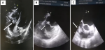
A 60-year-old diabetic man was admitted in cardiology department for dyspnea and fever evolving over 3 weeks with urinary symptoms. In his past history there was a COVID-19 infection that required hospitalization with oxygen therapy for a week and he reported repetitive urinary tract infections. The physical exam showed an axillary temperature at 39.5°c crackling sounds on the lung and no right ventricular failure signs. Laboratory tests showed biologic inflammatory markers elevation; cytobacteriological urine test isolated: enterococcus foecalis and several hemocultures isolated the same named bacteria. Transthoracic echocardiography showed a preserved left ventricular function, a vegetation at the expense of the anterior leaflet of mitral valve (7*4mm); mild mitral valve regurgitation and a huge vegetation at the expense of tricuspid valve (15*20mm) with important tricuspid regurgitation and pulmonary arterial hypertension. The transesophageal echocardiography showed the same vegetation at the expense of the mitral and tricuspid valve and no abnormalities in the other valves. After an initial antibiotic therapy, the patient was referred for surgery in front of worsening mitral insufficiency which has become grade 4.
Figure 1: A) huge vegetation at the expense of the tricuspid valve in the subcostal window (15*20mm); B) vegetation at the expense of the mitral valve in transesophageal echocardiography (7*4mm); C) vegetation at the expense of the tricuspid valve (15*20mm) in the transesophageal echocardiography (TEE)
Search
This article authors
On Pubmed
On Google Scholar
Citation [Download]
Navigate this article
Similar articles in
Key words
Article metrics
Recently from the PAMJ


Authors´ services
