
Kyste hydatique utérin: localisation rare
Issam Loukil, Amine Zouari
Corresponding author: Issam Loukil, Service de Chirurgie Générale Tataouine, Tataouine, Tunisie 
Received: 04 May 2021 - Accepted: 15 Aug 2021 - Published: 26 Aug 2021
Domain: General surgery
Keywords: Hydatidose, kyste, utérus
©Issam Loukil et al. Pan African Medical Journal (ISSN: 1937-8688). This is an Open Access article distributed under the terms of the Creative Commons Attribution International 4.0 License (https://creativecommons.org/licenses/by/4.0/), which permits unrestricted use, distribution, and reproduction in any medium, provided the original work is properly cited.
Cite this article: Issam Loukil et al. Kyste hydatique utérin: localisation rare. Pan African Medical Journal. 2021;39:272. [doi: 10.11604/pamj.2021.39.272.29631]
Available online at: https://www.panafrican-med-journal.com//content/article/39/272/full
Images in clinical medicine 

Kyste hydatique utérin: localisation rare
Kyste hydatique utérin: localisation rare
Hydatid cyst of the uterus: a rare localization
![]() Issam Loukil1,&, Amine Zouari2
Issam Loukil1,&, Amine Zouari2
&Auteur correspondant
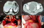
We here report the case of B.J, an 83-year-old patient with no previous history, presenting with intermittent abdominal pain evolving over the last few months. Physical examination revealed the presence of a firm, subumbilical mass that was tender to palpation. Ultrasound showed two multivesicular hydatid cysts (Gharbi�s classification type 3) located in segments III and IV of the liver and multilocular right latero-uterine cyst. Serologic test was positive. Tumor markers were negative. Abdomino-pelvic computed tomography (CT) scan showed two adjacent multivesicular hydatid cysts in the left side of the liver measuring 60x40 mm (A), and a multivesicular right latero-uterine pelvic hydatid cyst measuring 110x80 mm pushing the bladder forward and the uterus to the left (B). Surgical exploration revealed the presence of left hepatic cysts (C) and a huge hydatid cyst arising from the right edge of the uterine body (D). Resection of the salient domes was performed. All the precautionary measures were implemented to avoid possible peritoneal dissemination. Anatomopathological examination confirmed the diagnosis of hydatid cysts. One-year CT scan did not show any local or peritoneal recurrence.
Key words: hydatidosis, cyst, uterus

Patiente B.J âgée de 83 ans, sans antécédents, consulte pour des douleurs abdominales intermittentes évoluant depuis quelques mois. L´examen physique note la présence d´une masse abdominale sous ombilicale ferme et sensible à la palpation. Une exploration échographique a montré la présence de deux kystes hydatiques multi vésiculaires des segments III et IV du foie, type III selon la classification de Gharbi et un kyste latéro utérin droit multi loculé. La sérologie hydatique est positive. Les marqueurs tumoraux sont négatifs. Le scanner abdomino-pelvien a montré deux kystes hydatiques contigus du foie gauche multi vésiculaires de 60x40 mm (A), et un kyste hydatique pelvien latéro-utérin droit multivésiculaire de 110x80 mm refoulant la vessie en avant et l´utérus à gauche (B). L´exploration chirurgicale a mis en évidence les kystes du foie gauche (C) et un énorme kyste hydatique prenant naissance sur le bord droit du corps utérin (D). L´intervention chirurgicale par résection des dômes saillants a été réalisée avec toutes les mesures de précaution pour éviter une éventuelle dissémination péritonéale.
Figure 1: tomodensitométrie
(TDM) abdomino-pelvien: A) deux kystes hydatiques contigus du
foie gauche multivésiculaires de 60x40 mm; B) un kyste hydatique
pelvien latéro-utérin droit multivésiculaire de 110x80
mm refoulant la vessie en avant et l'utérus à gauche; C)
kystes du foie gauche; D) énorme kyste hydatique prenant naissance
sur le bord droit du corps utérin
Search
This article authors
On Pubmed
On Google Scholar
Citation [Download]
Navigate this article
Similar articles in
Key words
Article metrics
PlumX Metrics
Kyste hydatique utérin: localisation rareRecently from the PAMJ


Authors´ services
