
Dyke-Davidoff-Masson syndrome
Moli Jai Jain, Rakesh Krishna Kovela
Corresponding author: Rakesh Krishna Kovela, Department of Neuro-physiotherapy, Ravi Nair Physiotherapy College, Datta Meghe Institute of Medical Sciences, Wardha, Maharashtra, India 
Received: 30 Jul 2021 - Accepted: 13 Aug 2021 - Published: 20 Aug 2021
Domain: Radiology,Physical medicine and rehabilitation�or Physiatry,Neuroradiology
Keywords: Dyke-Davidoff-Masson syndrome, mirror movement, hand rehabilitation
©Moli Jai Jain et al. Pan African Medical Journal (ISSN: 1937-8688). This is an Open Access article distributed under the terms of the Creative Commons Attribution International 4.0 License (https://creativecommons.org/licenses/by/4.0/), which permits unrestricted use, distribution, and reproduction in any medium, provided the original work is properly cited.
Cite this article: Moli Jai Jain et al. Dyke-Davidoff-Masson syndrome. Pan African Medical Journal. 2021;39:256. [doi: 10.11604/pamj.2021.39.256.30993]
Available online at: https://www.panafrican-med-journal.com//content/article/39/256/full
Images in clinical medicine 

Dyke-Davidoff-Masson syndrome
Dyke-Davidoff-Masson syndrome
![]() Moli Jai Jain1, Rakesh Krishna Kovela2,&
Moli Jai Jain1, Rakesh Krishna Kovela2,&
&Corresponding author
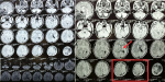
We are presenting a case of 15-year-old male patient presented with weakness of right upper and lower limbs, difficulty lifting objects with the right hand. He was born full term to non-consanguineous parents with no significant antenatal or perinatal history. On examination there was hemi-atrophy of the right side of the body with simultaneous involuntary movements of contralateral hand accompanying voluntary movement of the ipsilateral side. Neurological examination reveals right side spastic hemiparesis with upper limb more affected than lower limb. Tendon reflexes were brisk on affected side with extensor plantar response. Mini-mental status examination score was 26/30 suggestive of mild cognitive impairment. Other systemic examinations were within normal limits. Plain and contrast magnetic resonance imaging (MRI) of the brain findings, reveal left cerebral hemi-atrophy with thinning of the ipsilateral grey matter cortex, reduced volume of the underlying white matter, enlargement of the left lateral ventricle and reduced size of ipsilateral left cerebral peduncle is noted. Along with slight compensatory thickening of the ipsilateral skull vault is seen. Findings confirm diagnosis of Dyke-Davidoff-Masson Syndrome. He was under medical management along with physiotherapy rehabilitation focussing majorly on hand rehabilitation.
Figure 1: brain MRI findings shows left cerebral hemi-atrophy with thinning of the ipsilateral grey matter cortex, reduced volume of the underlying white matter (red box), enlargement of the left lateral ventricle (red arrow)
Search
This article authors
On Pubmed
On Google Scholar
Citation [Download]
Navigate this article
Similar articles in
Key words
Article metrics
PlumX Metrics
Dyke-Davidoff-Masson syndromeRecently from the PAMJ


Authors´ services
