
Rhino orbital mucormycosis
Shwetambari Morghade, Rakesh Krishna Kovela
Corresponding author: Rakesh Krishna Kovela, Department of Neuro Physiotherapy, Ravi Nair Physiotherapy College, Datta Meghe Institute of Medical Sciences, Sawangi Meghe, Wardha, Maharashtra, India 
Received: 28 May 2021 - Accepted: 02 Jun 2021 - Published: 09 Jun 2021
Domain: Pulmonology,Urgent Care Medicine,Neurology (general)
Keywords: Mucormycosis, COVID-19, intracranial extension, fungal infection
©Shwetambari Morghade et al. Pan African Medical Journal (ISSN: 1937-8688). This is an Open Access article distributed under the terms of the Creative Commons Attribution International 4.0 License (https://creativecommons.org/licenses/by/4.0/), which permits unrestricted use, distribution, and reproduction in any medium, provided the original work is properly cited.
Cite this article: Shwetambari Morghade et al. Rhino orbital mucormycosis. Pan African Medical Journal. 2021;39:114. [doi: 10.11604/pamj.2021.39.114.30053]
Available online at: https://www.panafrican-med-journal.com//content/article/39/114/full
Images in clinical medicine 

Rhino orbital mucormycosis
Rhino orbital mucormycosis
![]() Shwetambari Morghade1,
Shwetambari Morghade1, ![]() Rakesh Krishna Kovela1,&
Rakesh Krishna Kovela1,&
&Corresponding author

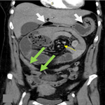
We are presenting the case of a 75-year-old female who tested positive for COVID-19 in the 2nd week of March with a high-resolution computed tomography (HRCT) score of 4/25 on the very next day for which she visited a hospital in sevagram where she was admitted for 7 days after which she was discharged and kept into home isolation under constant monitoring. After 2 months, on the 6th of May 2021 she was brought to the hospital in Sawangi, with complaints of pain over the right side of the face which was sudden in onset, continuous, dull aching, radiating to forehead on the right side with the history of associated swelling over the right side of the face which was initially small in size and gradually increased to present size of 3 x 2 cm approx, with history of difficulty in mastication, deglutition and speech were altered, nasal stiffness over the right side since 15 days. She is a known case of hypothyroidism for 20 years, diabetes mellitus and hypertension for 10 years with chronic kidney disease. On extraoral examination patient's face was asymmetrical due to fungal infection and diffused swelling over the right side of the face and opthalmoplegia, blurring of vision, ptosis, chemosis and restricted eye movements. Intraoral examination reveals mouth opening of 25mm with diffuse gingival swelling seen in the upper right maxillary alveolar region extending anteroposteriorly from 11 to 26 regions. Magnetic resonance imaging brain and orbit reveals invasive fungal sinusitis with the cutaneous collection and intracranial extension of mucormycosis.
Figure 1: (A,B,C) rhino orbital mucormycosis
Search
This article authors
On Pubmed
On Google Scholar
Citation [Download]
Navigate this article
Similar articles in
Key words
Tables and figures

Article metrics
PlumX Metrics
Rhino orbital mucormycosisRecently from the PAMJ


Authors´ services
