
Newborn with massive right eye proptosis revealing quintessence cystic orbital teratoma
Rohan Kumar Singh, Gaurav Vedprakash Mishra
Corresponding author: Rohan Kumar Singh, Department of Radiodiagnosis, Jawaharlal Nehru Medical College, Datta Meghe Institute of Medical Sciences, Sawangi (Meghe), Wardha, India 
Received: 26 May 2021 - Accepted: 29 May 2021 - Published: 07 Jun 2021
Domain: Radiology,Oncology,Pediatric ophthalmology
Keywords: Orbital teratoma, ultrasonography, orbital mass, neonate
©Rohan Kumar Singh et al. Pan African Medical Journal (ISSN: 1937-8688). This is an Open Access article distributed under the terms of the Creative Commons Attribution International 4.0 License (https://creativecommons.org/licenses/by/4.0/), which permits unrestricted use, distribution, and reproduction in any medium, provided the original work is properly cited.
Cite this article: Rohan Kumar Singh et al. Newborn with massive right eye proptosis revealing quintessence cystic orbital teratoma. Pan African Medical Journal. 2021;39:110. [doi: 10.11604/pamj.2021.39.110.30023]
Available online at: https://www.panafrican-med-journal.com//content/article/39/110/full
Images in clinical medicine 

Newborn with massive right eye proptosis revealing quintessence cystic orbital teratoma
Newborn with massive right eye proptosis revealing quintessence cystic orbital teratoma
Rohan Kumar Singh1,&, ![]() Gaurav Vedprakash Mishra1
Gaurav Vedprakash Mishra1
&Corresponding author

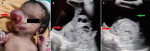
A full-term 2-day-old female neonate weighing about 2.17 kilogram, born by normal delivery at 37 weeks of gestation was referred to the ophthalmology with a massive mass of the right eye. On clinical examination, there was severe proptosis of the right eye with a positive transillumination test with normal left eye. The orbital mass was hard in consistency with few soft areas and it was covering the right midface. The eyeball was pushed on anteroinferior side with severe conjunctival chemosis and corneal erosion due to exposure to keratopathy with absent papillary reflex. For further routine investigation the baby was referred to the Department of Radiology for a B-scan ultrasound. On ultrasound, multiple anechoic cystic areas were seen anterior to globe pushing it anteroinferior, the hetero-echoic lesion was seen filling right eyeball and lesion was involving ocular muscles.
Figure 1: A) severe right eye proptosis with eyeball pushed out (red arrow); heterogeneous lesion with multiple anechoic cystic areas (red arrow); B) ultrasound images; C) heterogeneous well-defined lesion (red arrow), globe is pushed anteriorly (green arrow)
Search
This article authors
On Pubmed
On Google Scholar
Citation [Download]
Navigate this article
Similar articles in
Key words
Article metrics
Recently from the PAMJ


Authors´ services
