
Giant hepatic hemangioma: a challenging diagnosis
Sana Landolsi, Imène Ridène
Corresponding author: Sana Landolsi, Department of Surgery, Mahmoud El Matri Hospital, Ariana, Faculty of Medicine, University of Tunis El Manar, Tunis, Tunisia 
Received: 17 Aug 2020 - Accepted: 08 Apr 2021 - Published: 13 Apr 2021
Domain: Radiology,General surgery,Surgical oncology
Keywords: Giant hemangioma, cavernous liver hemangiomas, treatment
©Sana Landolsi et al. Pan African Medical Journal (ISSN: 1937-8688). This is an Open Access article distributed under the terms of the Creative Commons Attribution International 4.0 License (https://creativecommons.org/licenses/by/4.0/), which permits unrestricted use, distribution, and reproduction in any medium, provided the original work is properly cited.
Cite this article: Sana Landolsi et al. Giant hepatic hemangioma: a challenging diagnosis. Pan African Medical Journal. 2021;38:354. [doi: 10.11604/pamj.2021.38.354.25620]
Available online at: https://www.panafrican-med-journal.com//content/article/38/354/full
Images in clinical medicine 

Giant hepatic hemangioma: a challenging diagnosis
Giant hepatic hemangioma: a challenging diagnosis
Sana Landolsi1,&, Imène Ridène2
&Corresponding author

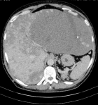
A 50-year-old man without medical history, complained about effort dyspnea with enlargement of the abdomen. Physical examination showed an enlarged liver reaching the right and the left iliac fossa with a collateral venous circulation. Biological exams were within normal ranges. Hepatitis B and C serologies were negative. Abdominal computed tomography and magnetic resonance imaging demonstrated an enlarged liver of 36 cm in diameter. Multiple heterogenous bilateral liver masses with enhancement after contrast intravenous injection were shown.The largest one with 26 cm in diameter was located in the left liver. A dense collateral venous circulation existed. A diagnostic percutaneous biopsy was made. Histopathological exam concluded to giant cavernous cavernoma. Since the surgery was risky especially hemorrhage, a percutaneous embolization for the largest mass was indicated.
Figure 1: giant cavernous hemangioma
Search
This article authors
On Pubmed
On Google Scholar
Citation [Download]
Navigate this article
Similar articles in
Key words
Tables and figures
Article metrics
PlumX Metrics
Giant hepatic hemangioma: a challenging diagnosisRecently from the PAMJ


Authors´ services
