
Unusual type of diabetes: fibrocalculous pancreatic diabetes
Natnicha Houngngam, Thiti Snabboon
Corresponding author: Thiti Snabboon, Excellence Center in Diabetes, Hormone and Metabolism, King Chulalongkorn Memorial Hospital, Thai Red Cross Society, Bangkok, Thailand 
Received: 01 Feb 2020 - Accepted: 20 Jan 2021 - Published: 03 Feb 2021
Domain: Endocrinology,Gastroenterology
Keywords: Fibrocalculous pancreatic diabetes, tropical chronic pancreatitis, ketosis-resistant diabetes
©Natnicha Houngngam et al. Pan African Medical Journal (ISSN: 1937-8688). This is an Open Access article distributed under the terms of the Creative Commons Attribution International 4.0 License (https://creativecommons.org/licenses/by/4.0/), which permits unrestricted use, distribution, and reproduction in any medium, provided the original work is properly cited.
Cite this article: Natnicha Houngngam et al. Unusual type of diabetes: fibrocalculous pancreatic diabetes. Pan African Medical Journal. 2021;38:116. [doi: 10.11604/pamj.2021.38.116.21663]
Available online at: https://www.panafrican-med-journal.com//content/article/38/116/full
Images in clinical medicine 

Unusual type of diabetes: fibrocalculous pancreatic diabetes
Unusual type of diabetes: fibrocalculous pancreatic diabetes
Natnicha Houngngam1, Thiti Snabboon1,2,&
&Corresponding author

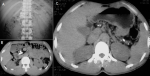
A 34-year-old Thai man complained about recurrent upper abdominal pain, polyuria and a 5kg weight loss in 6 months. He had no history of steatorrhea, jaundice, gallstones, alcohol intake, or cassava consumption. Physical examination was unremarkable except for his body mass index of 17.6 kg/m². The plasma glucose level was 672 mg/dL with normal serum amylase and lipase. Laboratory investigation showed serum osmolality of 287 mOsm/kg, bicarbonate 22 mEq/L and negative for serum ketone. Fibrocalculous pancreatic diabetes (FCPD) was diagnosed from a plain abdominal X-ray study showing multiple large calcifications over a pancreatic area (A). Computerized tomography of the abdomen revealed a large tubular calcification in dilated pancreatic duct during entering the duodenum (B). Scattered calcifications along the pancreatic duct were also noted (C). A genetic study revealed a SPINK1 N34S heterozygous mutation. His hyperglycemia responded well to insulin therapy. FCPD, a late stage of tropical chronic pancreatitis (TCP), is classified as a secondary cause of diabetes resulting from pancreatic dysfunction. The clinical picture consists of a triad of pancreatic calcification, abdominal pain, and diabetes. Its distinctive features are young age at onset, lean or underweight, ketosis-resistant diabetes, presence of large intraductal pancreatic calculi, and reported mainly in tropical developing countries. An etiology was previously thought to relate to malnutrition; however, a strong association with SPINK1 mutation is noted. The major morbidities are recurrent abdominal pain, steatorrhea, and malnutrition, while the majority of death is associated with diabetic nephropathy and pancreatic cancer. Treatment includes pancreatic enzyme supplementation, insulin therapy, and surgical drainage.
Figure 1: plain abdominal X-ray showed multiple large calcifications over pancreatic area (A), computerized tomography revealed a large tubular calcification in dilated pancreatic duct (B) and small scattered calcifications along pancreatic shadow (C)
Search
This article authors
On Pubmed
On Google Scholar
Citation [Download]
Navigate this article
Similar articles in
Key words
Article metrics
Recently from the PAMJ


Authors´ services
