
Pitfalls in spine injuries: risk of dramatic aortic dissection
Giuseppe Rosario Schirò, Pietro Domenico Giorgi
Corresponding author: Giuseppe Rosario Schirò, Grande Ospedale Metropolitano Niguarda, Milan, Italy 
Received: 31 Oct 2020 - Accepted: 11 Nov 2020 - Published: 16 Nov 2020
Domain: Radiology,Neurosurgery,Orthopedic surgery
Keywords: Spinal fracture, ankylosing spondylitis, trauma
©Giuseppe Rosario Schirò et al. Pan African Medical Journal (ISSN: 1937-8688). This is an Open Access article distributed under the terms of the Creative Commons Attribution International 4.0 License (https://creativecommons.org/licenses/by/4.0/), which permits unrestricted use, distribution, and reproduction in any medium, provided the original work is properly cited.
Cite this article: Giuseppe Rosario Schirò et al. Pitfalls in spine injuries: risk of dramatic aortic dissection. Pan African Medical Journal. 2020;37:242. [doi: 10.11604/pamj.2020.37.242.26771]
Available online at: https://www.panafrican-med-journal.com//content/article/37/242/full
Images in clinical medicine 

Pitfalls in spine injuries: risk of dramatic aortic dissection
Pitfalls in spine injuries: risk of dramatic aortic dissection
.gif) Giuseppe Rosario Schirò1,&, Pietro Domenico Giorgi1
Giuseppe Rosario Schirò1,&, Pietro Domenico Giorgi1
&Corresponding author

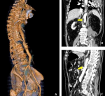
A 68 years old man was brought to the resuscitation room of our hospital following a fall from height. During the Focused Assessment with Sonography for Trauma-ultrasound (FAST-US), free liquid in abdomen was found. An angio-computed tomography (CT) (A, B, C) was performed showing a displaced T12 fracture in ankylosing spondylitis (AS) with a dangerous compression on the descending aorta by the displaced anterior wall of the fractured vertebra. This finding suggested high risk of iatrogenic aortic lesion during the surgical reduction of the fracture. Therefore, combined vascular and spinal surgery was deemed necessary. An inflatable balloon was placed endovascularly ready to stop the eventual bleeding and an endovascular prosthesis was ready to be positioned to repair the vascular lesion during the spine surgery. In this way, fracture reduction and fixation were safely performed. Aortic injury associated to thoracic spine fracture in AS is uncommon but potentially fatal. Due to the inflammation and adventitial scarring, the aorta can become tethered to the anterior longitudinal ligament and thus is subjected to shearing forces in an acute trauma. The fracture is generally associated to hyperextension mechanism, largely due to preexisting kyphotic deformity and there is a strong association with unstable B like-chance fractures. Undiagnosed vascular compression could be very dangerous leading to fatal dissection during spine surgery with mortality approaching 60%. In conclusion, spine injuries for vertebral fracture in patient with AS should be considered insidious and at risk of dramatic vascular lesion. Angio-CT and multidisciplinary management should be considered mandatory in cases of displaced fractures.
Figure 1: 3-D reconstruction showing aortic compression caused by the displaced fragments of an unusual spinal fracture pattern
Search
This article authors
On Pubmed
On Google Scholar
Citation [Download]
Navigate this article
Similar articles in
Key words
Tables and figures
Article metrics
Recently from the PAMJ


Authors´ services
