
Giant mediastinal mass
Danilo Coco, Silvana Leanza
Corresponding author: Danilo Coco, Department of General Surgery, Ospedali Riuniti Marche Nord, Pesaro, Italy 
Received: 29 Sep 2020 - Accepted: 10 Oct 2020 - Published: 15 Oct 2020
Domain: Thoracic surgery
Keywords: Giant mediastinal mass, mediastinal lymphoma B-cells, computed tomography
©Danilo Coco et al. Pan African Medical Journal (ISSN: 1937-8688). This is an Open Access article distributed under the terms of the Creative Commons Attribution International 4.0 License (https://creativecommons.org/licenses/by/4.0/), which permits unrestricted use, distribution, and reproduction in any medium, provided the original work is properly cited.
Cite this article: Danilo Coco et al. Giant mediastinal mass. Pan African Medical Journal. 2020;37:162. [doi: 10.11604/pamj.2020.37.162.26304]
Available online at: https://www.panafrican-med-journal.com//content/article/37/162/full
Images in clinical medicine 

Giant mediastinal mass
Giant mediastinal mass
Danilo Coco1,&, Silvana Leanza2
&Corresponding author

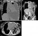
A 43-year-old Caucasian female presented to the emergency department (ED) with significant dyspnea, thoracic pain and fever. She presented a negative medical history and no therapy. During the physical examination, the patient was uncomfortable. Her vital signs were: blood pressure, 100/90 mmHg; respiratory rate, 50 breaths/minute; heart rate, 130 beats/minute; and temperature superior to 38°C. Oxygen saturation was 80% on room air and 90% with the aid of oxygen. The abdominal examination was unremarkable. Laboratory evaluation revealed high leukocytosis with a white blood cell (WBC) count of 15 per mm3. Arterial Blood Gases (ABG) demonstrated respiratory acidosis: PO2 80, PCO2 60, HCO3 30 mEq. Thoracic X-ray revealed a massive pleural effusion. Computed tomography demonstrated a giant mediastinal mass surrounding pulmonary artery, aorta and pericardia pleura associated with massive pleural effusion. The patients immediately started intravenous (IV) fluids of 2l in 6 hours, Foley and jugular catheter vein cannulation to support main arterial pressure and urine output. The patient was transferred to surgical services where a 28 Fr thoracic drainage was inserted. Post-drainage thoracic scan (CT) demonstrated only the giant mediastinal mass. Fine-needle aspiration (FNA) CT scan guided was performed. Histopathological findings were mediastinal lymphoma B-cells. The patient was discharged three days after.
Figure 1: A) thoracic computed tomography (CT) scan demonstrating a giant mediastinal mass associated with massive left pleural effusion; B) post-28 Fr thoracic drainage demonstrating the extension of giant mediastinal mass; C) FNA thoracic CT scan biopsy
Search
This article authors
On Pubmed
On Google Scholar
Citation [Download]
Navigate this article
Similar articles in
Key words
Article metrics
PlumX Metrics
Giant mediastinal massRecently from the PAMJ


Authors´ services
