
A rare femoral tumor in a young patient
Faten Limaiem
Corresponding author: Faten Limaiem, University of Tunis El Manar, Tunis Faculty of Medicine, 1007, Tunisia 
Received: 05 Apr 2020 - Accepted: 10 Apr 2020 - Published: 07 Oct 2020
Domain: Oncology,Orthopedic surgery
Keywords: Parosteal osteosarcoma, bone, tumor, pathology
©Faten Limaiem et al. Pan African Medical Journal (ISSN: 1937-8688). This is an Open Access article distributed under the terms of the Creative Commons Attribution International 4.0 License (https://creativecommons.org/licenses/by/4.0/), which permits unrestricted use, distribution, and reproduction in any medium, provided the original work is properly cited.
Cite this article: Faten Limaiem et al. A rare femoral tumor in a young patient. Pan African Medical Journal. 2020;37:135. [doi: 10.11604/pamj.2020.37.135.22667]
Available online at: https://www.panafrican-med-journal.com//content/article/37/135/full
Images in clinical medicine 

A rare femoral tumor in a young patient
A rare femoral tumor in a young patient
Faten Limaiem1,&
&Corresponding author

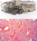
Parosteal osteosarcoma is a low-grade, bone-forming neoplasm that arises on the surface of bone. It accounts for about 4% of all osteosarcomas. An 18-year-old male patient with no particular past medical history, consulted for a painless mass in the right thigh that had appeared at the age of 17 years and progressively increased in volume. The physical examination revealed a 6 cm mass at its largest above the right popliteal fossa with knee flexion slightly limited. The X-ray revealed a well-limited mass in the lower third of the femur that was dense and attached to the metaphyseal cortex by a wide base. Histological examination of the biopsy specimen established the diagnosis of parosteal osteosarcoma. The patient underwent wide resection of the femoral tumor (A) preceded by a course of first-line chemotherapy. Histological examination showed a malignant mesenchymal proliferation, moderately cellular, made up of long, linear and eosinophilic material, sometimes calcified with no osteoblastic cells in the periphery (B). The tumor cells were spindle-shaped, with little eosinophilic cytoplasm and a long or ovoid, hyperchromatic, and moderately atypical nucleus. Mitoses were rare. There were no areas of dedifferentiation. Postoperative course was uneventful. During the one-year follow-up period, there was no recurrence or metastasis of the tumor. Parosteal osteosarcoma is characterized by its insidious growth and favorable prognosis. It rarely leads to metastasis. Its treatment is mainly surgical.
Figure 1: (A) macroscopic examination of the surgical specimen showing a lobulated, whitish tumor with focal cartilaginous zones measuring 6.3 cm x 2.8 cm, attached to the bone by a wide base; (B) photomicrograph of parosteal osteosarcoma showing mature-appearing bone without osteoblastic rimming, surrounded by a hypercellular fibroblastic stroma with moderate cytologic atypia, magnification (x200)
Search
This article authors
On Pubmed
On Google Scholar
Citation [Download]
Navigate this article
Similar articles in
Key words
Article metrics
PlumX Metrics
A rare femoral tumor in a young patientRecently from the PAMJ


Authors´ services
