
Tracheobronchial calcifications in a child
Houda Rahmoun, Chafiq Mahraoui
Corresponding author: Houda Rahmoun, Department of Pediatrics I, Children´s Hospital of Rabat, Rabat, Morocco 
Received: 05 Aug 2020 - Accepted: 07 Aug 2020 - Published: 24 Aug 2020
Domain:
Warning: Use of undefined constant ARTDOMAIN - assumed 'ARTDOMAIN' (this will throw an Error in a future version of PHP) in /home/panafric/panafrican-med-journal.com/content/article/index_article.php on line 296
ARTDOMAIN
Keywords: Tracheobronchial calcifications, Keutel syndrome, child
©Houda Rahmoun et al. Pan African Medical Journal (ISSN: 1937-8688). This is an Open Access article distributed under the terms of the Creative Commons Attribution International 4.0 License (https://creativecommons.org/licenses/by/4.0/), which permits unrestricted use, distribution, and reproduction in any medium, provided the original work is properly cited.
Cite this article: Houda Rahmoun et al. Tracheobronchial calcifications in a child. Pan African Medical Journal. 2020;36:331. [doi: 10.11604/pamj.2020.36.331.25384]
Available online at: https://www.panafrican-med-journal.com//content/article/36/331/full
Images in clinical medicine 

Tracheobronchial calcifications in a child
Tracheobronchial calcifications in a child
Houda Rahmoun1,&, Chafiq Mahraoui1
&Corresponding author

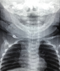
Three months old child was admitted in the department of pediatrics I for respiratory distress, fever and laryngeal stridor. He was born to consanguineous parents. The prenatal history was unremarkable. However, he was hospitalized at birth for respiratory distress which improved quickly with oxygen nasal cannula. Since one month old, he was presenting a laryngeal stridor, persistent cough and chest congestion. Clinical examination found a dyspnea, stridor occuring in both phases of respiration, signs of retractions, a dysmorphic face with mid-facial hypoplasia and brachytelephalengia. The chest x-ray showed calcifications involving the entire tracheobronchial tree. A chest computed tomography revealed bilateral and symmetrical calcifications involving the anterior cartilaginous part of the trachea and the stem bronchi. The transthoracic ultrasound was normal. Routine physicochemical examinations found a low prothrombin time with decrease in the levels of vitamin-k dependent coagulation factors. The clinical course was favorable with oxygen nasal cannula and respiratory physiotherapy. The diagnosis of Keutel syndrome was made on calcifications of the tracheal cartilage associated to brachytelephalengia, and also facial dysmorphism.
Figure 1: tracheobronchial calcifications
Search
This article authors
On Pubmed
On Google Scholar
Citation [Download]
Navigate this article
Similar articles in
Key words
Tables and figures
Article metrics
PlumX Metrics
Tracheobronchial calcifications in a childRecently from the PAMJ


Authors´ services
